Uveitis: When the Inside of the Eye Is Inflamed
Claire Greenway
BVM&S MRCVS
You may have arrived here from the red-eye page, or with a diagnosis that surprised you, because what started as "a sore eye" has turned into a conversation about blood tests. That can feel like a swerve, so here's why it isn't.
Uveitis is inflammation inside the eye, and the eye is one of the few places a vet can look straight at living blood vessels. So when those vessels inflame, the eye is sometimes the first part of your pet to signal that something is going on more widely. The blood tests aren't over-testing. They're the eye doing its job as an early-warning window onto the whole animal, and that idea is the spine of everything below. (If you're still at the "how worried should I be" stage, the urgency sorting belongs on the red, painful eye page; this page picks up the inside-the-eye story.)
What "uveitis" actually means
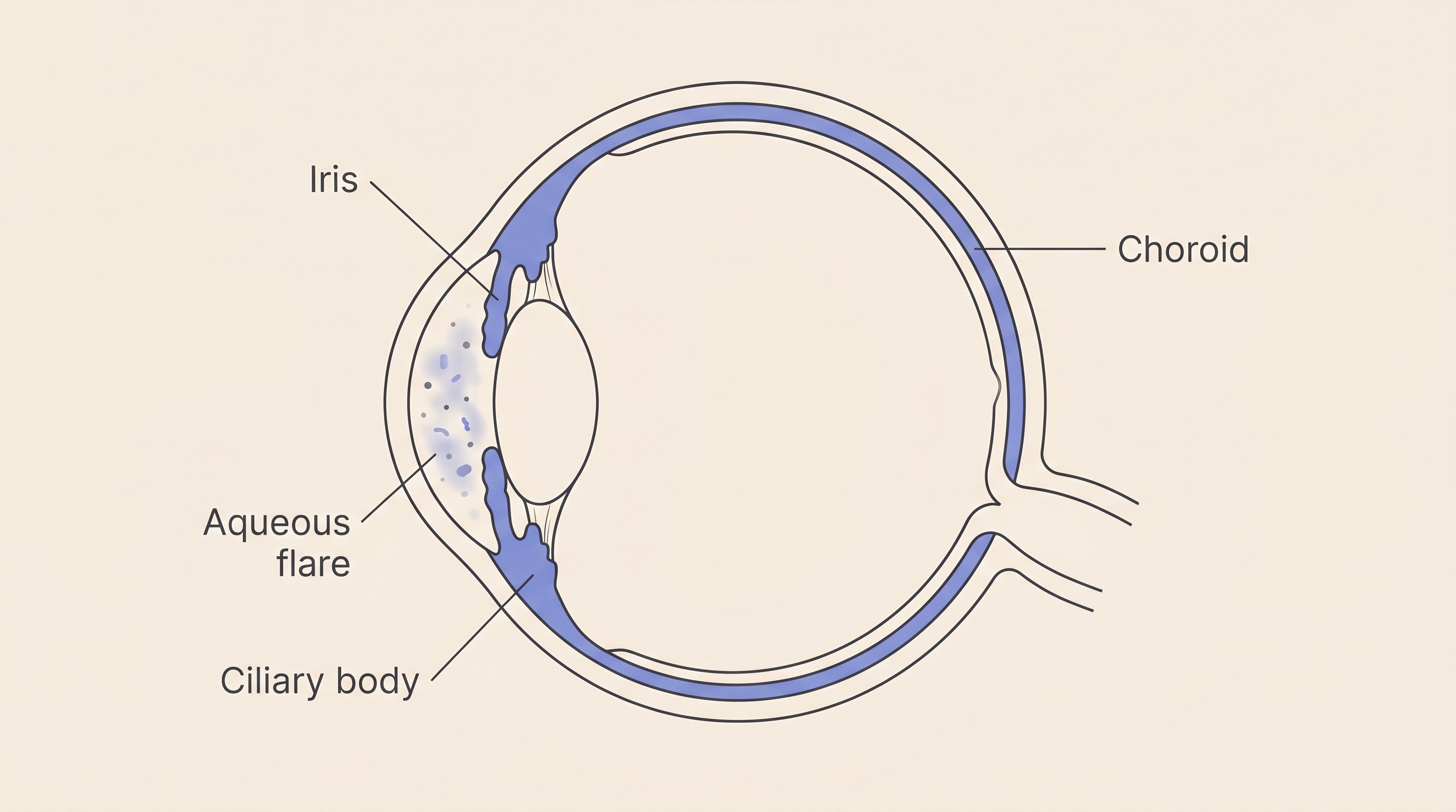
The technical bit first, said once. Uveitis is inflammation of the uvea, "the highly vascular and often pigmented iris, ciliary body, and choroid" (Townsend, 2008): the eye's middle layer, its plumbing and blood-supply layer. When it inflames it leaks, releasing cells, protein and sometimes blood into the chambers that are meant to be crystal clear, which is what makes a uveitic eye look hazy or bloodshot and is part of why it hurts (Townsend, 2008).
The sub-types just describe where it sits: anterior uveitis (iridocyclitis) at the front, posterior uveitis (chorioretinitis) at the back, and panuveitis when the whole uvea is involved (Townsend, 2008; Today's Veterinary Practice, 2019). That matters more than it sounds, because drops reach the front of the eye but not the back, so posterior disease needs medicine by mouth (Townsend, 2008).
The headline: uveitis is a symptom, not a diagnosis
This is the message I most want to land. Uveitis is not one disease. It's "a non-specific response to many different underlying causes, many of which are systemic diseases (inside of the body)" (ACVO, n.d.). The eye can only inflame in a limited number of ways, so many separate problems funnel into the same red, sore, hazy picture.
Vets sort the causes into five buckets: immune-mediated, lens-induced, infectious, secondary to cancer and traumatic (ACVO, n.d.). The biggest is the immune-mediated one, roughly 75% of cases, and it "is a diagnosis of exclusion, because there is no specific test to diagnose immune-mediated uveitis" (ACVO, n.d.). We reach that label only by testing hard for the causes we can name and finding them clear, which is why the ACVO notes that "approximately 75% of cases fail to obtain a definitive diagnosis" (ACVO, n.d.). So an "idiopathic" verdict isn't the vet giving up. It means the system worked: we ruled out the treatable and the sinister, and can treat the inflammation itself with a clear conscience.
One urgent practical, because it's load-bearing for uveitis. A red, sore eye always earns a prompt vet look, today if it's very painful, and the one thing never to do is reach into the cupboard for an old steroid eye drop. Topical steroids are a mainstay for immune-mediated uveitis, but they are "contraindicated with corneal ulceration or infectious keratitis" because they delay healing and worsen infection (Today's Veterinary Practice, 2019). Since you can't tell at home whether that red eye has an ulcer hiding in it, a cupboard steroid can turn a treatable scratch into a disaster. Our Eye-emergency triage tool helps you judge how fast to be seen.
Why the blood tests: the eye as a window
Because so many whole-body diseases can surface as uveitis, the work-up is deliberately broad: "a thorough physical examination, complete blood cell count, serum biochemistry profile, and urinalysis should be performed" (Townsend, 2008), usually with targeted blood tests aimed at the likely culprits, and sometimes chest x-rays or a scan to hunt for cancer or deep infection (Today's Veterinary Practice, 2019; ACVO, n.d.). What we test for diverges between dogs and cats, and that divergence is part of the story.
In dogs, the biggest bucket has no nameable cause: in the canine reference series, "60% of cases of dogs that had uveitis were classified as idiopathic or immune mediated" (Townsend, 2008). The rest spreads across bloodborne infections (tick-borne disease, Brucella, leptospirosis and the systemic fungal infections, which depend on where a dog lives and travels), alongside trauma and cancer (Townsend, 2008; Today's Veterinary Practice, 2019). Two dog causes deserve a name, since this page owns the round-up. Golden Retriever pigmentary uveitis (GRPU) is a common, breed-specific, late-life inflammation, marked by radial flecks of pigment on the front of the lens, that can progress quietly to glaucoma and blindness: in the Purdue follow-up series the share of affected eyes still holding vision fell from 98.3% to 84.2%, with posterior synechiae and fibrin in the front chamber the significant risk factors for glaucoma, and almost half of affected dogs lose vision in an eye within a year of diagnosis; the ACVO Genetics Committee recommends affected dogs are not bred (Jost et al., 2020). Uveodermatologic syndrome (a VKH-like syndrome) is the other: an immune attack on the body's pigment cells, so uveitis arrives alongside whitening of the nose, lips and coat, classically in Akitas, needing long-term immunosuppression (Townsend, 2008). Sometimes the skin gives the game away before the eye does.
In cats, the "look further" message earns its keep, because infectious and systemic causes loom much larger. A meaningful share is still idiopathic, with no cause found (Townsend, 2008; Jinks et al., 2016), but the rest is dominated by a recognisable set of infections worth testing for. In a referral series of 120 cats with uveitis, the agents tested for showed up at notable rates: Bartonella in 43.2%, feline coronavirus in 34.7%, Toxoplasma in 23.7%, FIV in 7.3% and FeLV in 2.7%, with at least one agent found in 59.2%; feline infectious peritonitis was diagnosed in 15.8% and cancer in 5.0%, while 40.8% had no cause identified (Jinks et al., 2016). Add the systemic fungal infections, especially cryptococcosis, and that is the feline panel (Lappin, 2000; Townsend, 2008).
A caveat keeps those honest: they come from US referral clinics, so they show which infections to test for, not how common each is in a British cat, and antibodies show exposure rather than proof that agent caused this cat's uveitis (Bartonella, for one, is better thought of as "tested for" than "to blame") (Jinks et al., 2016; Lappin, 2000). The takeaway holds regardless: in a cat, uveitis is a flag to go looking, because finding the cause can change the outcome, and because some of what we find changes the bigger picture for the whole cat, not just the eye (Jinks et al., 2016; Lappin, 2000).
The lens that turns on the eye
One more cause ties this page to the cataract section, and it surprises people: a cataract isn't an inert grey disc. A mature or hypermature cataract leaks lens protein, and because the immune system never normally "sees" the inside of the lens, it treats that escaping protein as foreign and attacks it (Townsend, 2008). Leakage through an intact capsule is phacolytic uveitis, the milder grumble from an advancing cataract; a frank rupture of the capsule, which can happen when a diabetic cataract swells fast, is phacoclastic uveitis, the more severe one (in the classic pathology study of lens-capsule rupture in dogs, proliferating lens tissue, a sealed-over pupil and glaucoma affected 11 of the 20 eyes) (Wilcock and Peiffer, 1987). This is why an advancing cataract needs watching, and why diabetic dogs (around 80% of whom develop cataracts within the first year or so) are referred early (Beam et al., 1999). What to do about the cataract itself belongs to cataracts explained and living with cataracts without surgery.
The complications, and why "treat it properly" matters
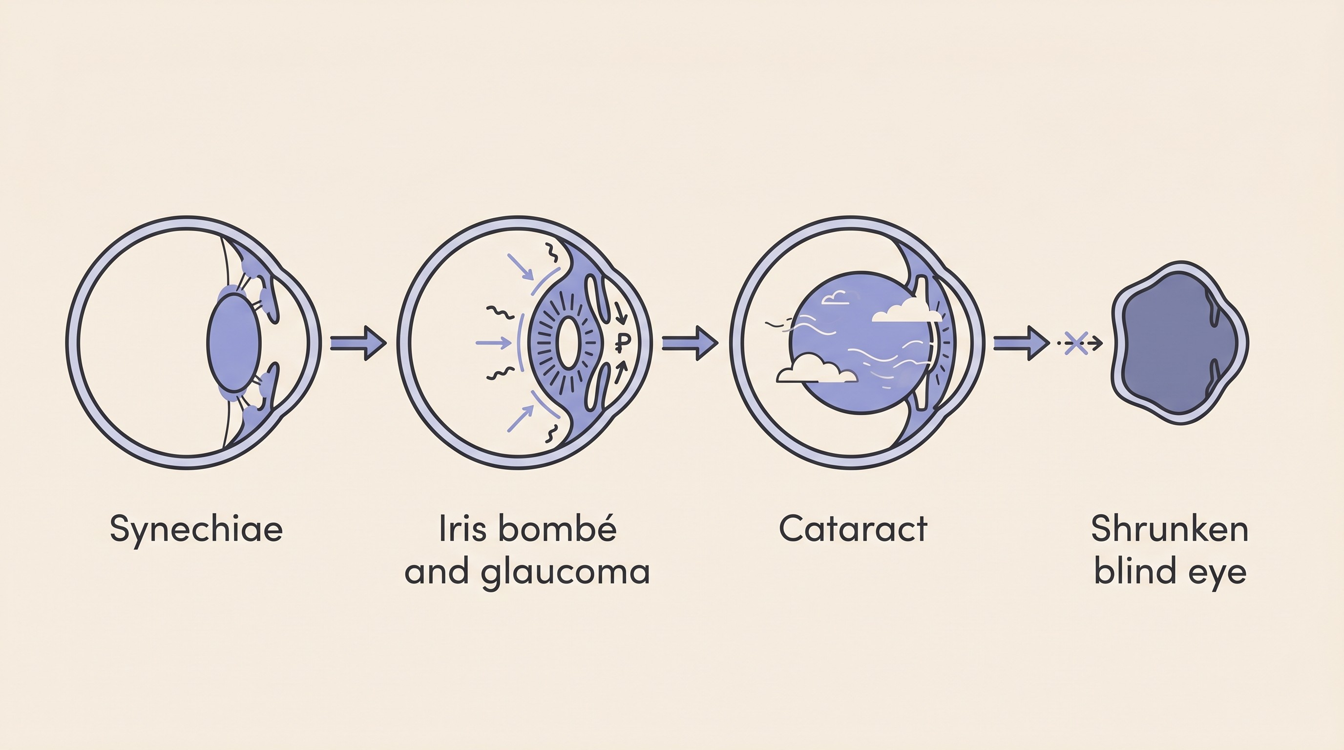
Uveitis is treated aggressively because, left to smoulder, it has a handful of ways to cost an eye. The most feared is secondary glaucoma. Inflammation makes the iris stick to the lens behind it, forming scars (posterior synechiae); if that scarring runs all the way around the pupil, fluid is trapped behind the iris, which balloons forward (iris bombé) and blocks the eye's drainage, so the pressure climbs (Townsend, 2008; Today's Veterinary Practice, 2019). This matters especially in cats, where chronic uveitis is the leading route into glaucoma, so I'll hand the rest to glaucoma in dogs and glaucoma in cats. Chronic inflammation also drives cataract, weakens the fibres that hold the lens (a frequent cause of lens slippage in cats), can detach the retina, and at the end of the road can leave an eye shrunken and blind, a state called phthisis bulbi (Townsend, 2008; ACVO, n.d.). The ACVO's blunt line is the takeaway: "Uveitis must be treated aggressively in order to prevent complications that lead to blindness" (ACVO, n.d.). The feline secondary cataract that shares this root cause is covered in cataracts in cats.
One counter-intuitive sign is worth knowing, because it's how a flare and its worst complication are told apart. In uveitis the pressure inside the eye usually falls, because the inflamed ciliary body makes less fluid, so a low reading points to uveitis while a rising one is the alarm that it has tipped into secondary glaucoma (Townsend, 2008). It's why your vet checks the pressure rather than judging by eye, and why a uveitic eye that suddenly turns harder, more painful or more clouded needs to be seen promptly.
Treatment: calm the inflammation, and treat the cause
Treatment runs on two tracks at once. The first is calming the inflammation, with the goals of "halting inflammation, stabilizing the blood-aqueous barrier, minimizing sequelae, decreasing pain, and preserving vision" (Townsend, 2008). That usually means an anti-inflammatory eye drop, a topical corticosteroid such as prednisolone acetate (or a topical NSAID where a steroid isn't safe), often paired with a dilating drop (atropine) to ease the painful spasm of the inflamed ciliary muscle and help stop the iris scarring onto the lens (Townsend, 2008; Today's Veterinary Practice, 2019). When disease is at the back of the eye or severe, medicine by mouth is added, because "topical treatments do not benefit the posterior segment" (Townsend, 2008).
The second track is treating any underlying cause: "Appropriate treatment of underlying disease (when possible) is paramount, as insufficient management will result in recurrent or persistent uveitis" (Today's Veterinary Practice, 2019), which might mean antibiotics or antifungals for infection, or immunosuppressants such as azathioprine or ciclosporin for stubborn immune-mediated disease. Whatever the regime, it's tapered slowly, not stopped the moment the eye looks better: treatment generally continues for "2 to 4 weeks past the resolution of clinical signs" (Today's Veterinary Practice, 2019), because pulling the medicine too early is one of the surest ways to relight the fire.
A few drops a day, sometimes per eye and sometimes ongoing, is a real commitment, and missed doses are where well-controlled eyes slip. The eye-drop and pressure tracker carries the timing and reminders, and managing glaucoma at home covers long-term dosing, which applies just as neatly to a uveitis routine. One honest note: if uncontrolled uveitis has already blinded an eye and left it painful, removing that eye takes away constant pain and is one of the kindest operations we do, not a failure (enucleation and eye removal covers that decision).
That's the shape of it. Take a red, sore eye to be seen promptly, never guess from the cupboard, let your vet run the bloods, and take "no cause found" as the normal, reassuring answer it is. Treated properly and tapered patiently, most uveitic eyes settle and do well: the job is to calm the inflammation, find and treat anything underneath it, and keep half an eye on the pressure. If you'd like to get better at reading the quiet signs of a sore eye before it flares, spotting eye pain is worth having in your back pocket.
References
- American College of Veterinary Ophthalmologists (ACVO). Uveitis in Dogs and Cats. ACVO Public Resources, n.d.
- Beam, S., Correa, M. T., & Davidson, M. G. (1999). A retrospective-cohort study on the development of cataracts in dogs with diabetes mellitus: 200 cases. Veterinary Ophthalmology, 2(3), 169-172.
- Jinks, M. R., English, R. V., & Gilger, B. C. (2016). Causes of endogenous uveitis in cats presented to referral clinics in North Carolina. Veterinary Ophthalmology, 19(Suppl 1), 30-37.
- Jost, H. E., Townsend, W. M., Moore, G. E., & Liang, S. (2020). Golden retriever pigmentary uveitis: Vision loss, risk factors for glaucoma, and effect of treatment on disease progression. Veterinary Ophthalmology, 23(6), 1001-1008.
- Lappin, M. R. (2000). Feline infectious uveitis. Journal of Feline Medicine and Surgery, 2(3), 159-163.
- Today's Veterinary Practice. (2019). Managing uveitis in dogs and cats.
- Townsend, W. M. (2008). Canine and feline uveitis. Veterinary Clinics of North America: Small Animal Practice, 38(2), 323-346.
- Wilcock, B. P., & Peiffer, R. L. (1987). The pathology of lens-induced uveitis in dogs. Veterinary Pathology, 24(6), 549-553.
Keep track of how your pet is doing
The owners who cope best are the ones who notice changes early. A simple health log shows you what is working, and what is not, before the next vet visit.
Start tracking, freeYou're not doing this alone
Compare treatment journeys and talk to owners managing vision & eye health. Free to join.
Join PetsLikeMine