Glaucoma in Dogs: The Painful Emergency Owners Miss

Glaucoma in Dogs: The Painful Emergency Owners Miss

D

Dr. Alastair Greenway

MRCVS

14 Jun 202614 min read0 views
Vet reviewedby Claire Greenway, BVM&S MRCVSLast reviewed 13 Jun 2026

I'll start with the part I most need you to hear, because everything else here can wait two minutes and this cannot. If your dog has an eye that's suddenly turned red, cloudy or hazy, that looks bigger or more bloodshot than the other one, that they're squinting or rubbing, or that seems to have stopped seeing, ring your vet now and say you think it might be glaucoma. Today, not tomorrow morning. Acute glaucoma is an ophthalmological emergency (Thomasy & Carnevale, 2024), and it's one of those rare eye problems where keeping the sight or losing it can come down to hours. If you want a quick way to gauge how urgent the signs in front of you are, our Eye-emergency triage walks you through it, and the eye-emergency red-flags sheet is worth keeping on the fridge.

Now, with the phone hopefully ringing, let me explain what's going on, why it hurts far more than most owners realise, and why dogs are so good at hiding it that it slips past loving, attentive people all the time.

Flat illustration of a dog's eye in cross-section showing aqueous fluid building up behind a blocked drainage angle, pressing on the retina and optic nerve at the back
Fluid is made inside the eye all the time and normally drains away. In glaucoma the drain is blocked, so pressure climbs and presses on the retina and optic nerve.

What glaucoma actually is

The eye makes a clear fluid called aqueous humour all the time, and in a healthy eye it drains away just as fast as it's produced, keeping the internal pressure steady. Glaucoma is what happens when the drainage falls behind: fluid keeps arriving, can't get out fast enough, and the pressure climbs. That raised pressure is the whole problem, because it crushes the delicate retina and strangles the blood supply to the optic nerve, the cable that carries vision to the brain (Cornell Riney; Komáromy et al., 2019). It's best understood not as one disease but as a group of conditions that all end the same way: optic nerve damage, death of the retinal nerve cells, and irreversible blindness (Komáromy et al., 2019).

For a sense of scale, normal pressure inside a dog's eye sits around 15 to 25 mm Hg (Reinstein, 2018). In a full acute attack it can rocket past 40 to 60 mm Hg (Thomasy & Carnevale, 2024). That's the eye being inflated like an over-pumped tyre, and the tissues inside it aren't built to take it. You can't read that pressure by looking, which is the first reason this is a vet's job: the diagnosis turns on a number from a little instrument called a tonometer rested gently on the eye.

It hurts. Really hurts.

This is the part owners almost never know, and it's the part I most want to land. A high-pressure glaucoma attack is genuinely, severely painful. In people the same rise in eye pressure causes a headache ranging from a bad migraine to something that brings on nausea and vomiting, and dogs have the very same pain machinery in their eyes (Texas A&M, Pet Talk). The veterinary literature describes glaucoma, plainly, as "an often painful, complex group of blinding optic neuropathies" (Komáromy et al., 2019). The pain tracks the pressure: the higher it climbs, the worse it hurts, and the only thing that truly relieves it is bringing the pressure down (Miller, Canine Glaucoma).

So why do so many owners miss it? Because dogs don't show pain the way we expect. Some squint, hold the eye shut or paw at it, but many simply mask it: they go quiet, sleep more, seem flat and off their food, and read as "a bit off" or "just getting old" (Texas A&M, Pet Talk; Reinstein, 2018). Lethargy, a smaller appetite and sleeping more are exactly the quiet pain signals that get overlooked. The clue is often the change afterwards, because once the pressure is treated owners frequently describe a dog who is suddenly brighter and more themselves again (Texas A&M, Pet Talk). That before-and-after tells you the eye had been hurting all along.

Reading those quiet signs is a skill worth having across every eye condition, not just this one, and my colleague has written that piece: spotting eye pain.

The signs you can actually see

Two-column check card contrasting the visible signs of a glaucoma attack with the quiet behaviour changes owners miss
A glaucoma attack shows up two ways at once: the eye you can see and the dog you can feel. A red, cloudy eye plus a quiet, flat dog is a today emergency.

In an acute attack, alongside that quiet behaviour change, there are physical signs you can spot at home:

  • A red, angry-looking eye. The redness of glaucoma is often the deep, branching vessels on the white of the eye standing up and engorged, rather than the pink, surface fizz of simple conjunctivitis, though you don't need to tell those apart yourself (Miller, Canine Glaucoma).
  • A bluish or cloudy haze across the front of the eye, which is the cornea swelling with fluid (Thomasy & Carnevale, 2024; Cornell Riney).
  • Squinting, blinking or holding the eye partly shut (blepharospasm), and sometimes a watery eye (Reinstein, 2018).
  • A pupil that's larger than the other side and doesn't shrink much in bright light (Miller, Acute Primary Angle-Closure Glaucoma).
  • The eye appearing blind: not tracking movement, bumping into things on that side (Cornell Riney).

A bulging, visibly enlarged eyeball, which vets call buphthalmos, tends to come later. It signals the eye has been under high pressure for a while, so it usually points to a more chronic stage than the first hours of an attack (Thomasy & Carnevale, 2024). If you're already seeing a stretched, enlarged globe, that doesn't mean it's hopeless, but it does mean don't wait any longer.

A useful rule while you decide: you don't have to be sure it's glaucoma, and you can't be from the sofa. A red, cloudy or painful eye, or one that's clearly stopped seeing, earns a same-day call whatever the cause. The triage tool exists for exactly this "is this an emergency or am I overreacting" moment.

Why hours genuinely matter

I want to be honest here, because there's a difference between urgency and false hope, and you deserve the real version.

Speed matters because the nerve cells in the retina start dying quickly under high pressure, and there's a cruel twist: as those cells die they release chemicals that go on to kill their healthy neighbours, a chain reaction that can keep rolling even after the pressure is back to normal (Miller, Acute Primary Angle-Closure Glaucoma). The practical salvage window is short, often less than 12 to 24 hours (Reinstein, 2018). After that the odds drop fast. If the pressure has sat above 50 mm Hg for more than three days the chance of saving useful vision is essentially gone, and an eye holding above 40 mm Hg will very likely end up blind (Miller, Canine Glaucoma).

And even when a dog is rushed in and treated promptly during an attack, only around half of eyes regain vision, partly because the pressure has often been spiking quietly for a while before the dramatic attack that finally gets noticed (Reinstein, 2018). So the message isn't "rush in and we can always fix it". It's "get seen fast, because that's how we find out which eye can still be saved, save what we can, and stop the pain either way". On the days we can't rescue the sight, we can always rescue your dog from the pain, and that matters enormously.

Primary versus secondary: the fork that decides what comes next

Glaucoma comes in two broad types, and which one your dog has changes almost everything afterwards, including the fate of the other eye.

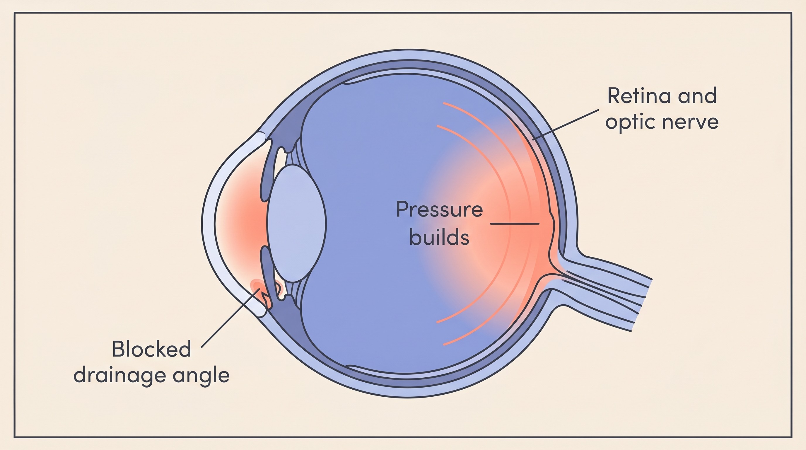
Primary glaucoma is an inherited fault in the eye's drainage system: the drainage angle is malformed (vets call this goniodysgenesis) or too narrow, and it eventually clogs (Reinstein, 2018; Cornell Riney). It's fundamentally a both-eyes disease that simply shows up in one eye first, because the drainage fault is built into the dog and is there in the other eye too.

Secondary glaucoma is when the drainage gets blocked by some other problem inside the eye, most often a lens that has slipped out of position (anterior lens luxation), long-standing inflammation (uveitis), an advanced cataract, bleeding inside the eye, a detached retina or a tumour (Miller, Canine Glaucoma; Reinstein, 2018). Here the glaucoma is a complication, so the underlying cause matters as much as the pressure. If uveitis is the driver, that inflammation needs treating in its own right.

That fork matters most because of the second eye, which I'll come to shortly.

This is common, and some breeds are far more at risk

Glaucoma isn't a rare curiosity. Counting primary and secondary together it affects roughly 1.7% of dogs in North America (Merck Veterinary Manual). The inherited primary form alone has been climbing in purebred dogs over the decades, reaching 0.89% by the late 1990s and early 2000s (Gelatt & MacKay, 2004).

It's also strongly breed-linked. In the largest dataset the highest-risk breeds were the American Cocker Spaniel (5.52%), Basset Hound (5.44%), Chow Chow (4.70%), Shar-Pei (4.40%), Boston Terrier (2.88%) and Wire Fox Terrier (2.28%) (Gelatt & MacKay, 2004). The Arctic breeds carry real risk too, the Siberian Husky, Akita and Samoyed among them, along with the Springer Spaniels, Great Dane, Flat-Coated Retriever, Beagle, Cairn Terrier and Miniature Poodle (Willows; Miller, Acute Primary Angle-Closure Glaucoma; Gelatt & MacKay, 2004). Onset is usually middle age, around four to nine years, and in several of these breeds females are affected noticeably more often than males, by roughly two to one in the angle-closure form (Miller, Acute Primary Angle-Closure Glaucoma; Gelatt & MacKay, 2004).

If you own one of these breeds this isn't a reason to lie awake. It's a reason to know the emergency signs cold, so that if an eye ever turns red and cloudy you act in hours rather than puzzling over it for a day. Awareness is the whole advantage. It's also worth asking your vet whether your breed is one where a baseline eye check or a gonioscopy (a look at the drainage angle) is worthwhile, because spotting a narrow angle before it ever shuts is the best position to be in.

The other eye is on a clock

This follows straight from the primary-versus-secondary fork, and it's one of the most important things to grasp if your dog has just been diagnosed with primary glaucoma in one eye. The second eye is genuinely at risk: in primary glaucoma there's about a 50% chance of an attack in the other eye within roughly 8 months if nothing is done (Miller et al., 2000; Cornell Riney). The good news, and it's real, is that preventive pressure-lowering drops can roughly halve that risk and buy a lot more time (Miller et al., 2000).

That makes protecting the fellow eye an active, worthwhile plan rather than waiting for the axe to fall, and it's important enough to have its own page. The protocol, the monitoring and the honest numbers all live in glaucoma in the fellow eye. With secondary glaucoma the picture differs: it's about whatever caused it, not an inevitable second attack on a built-in fault.

What treatment looks like, briefly

Emergency treatment is entirely a job for your vet or an out-of-hours clinic, and there's an important safety reason for that. The goal in the acute phase is to drop the pressure fast. The usual first move is a drop called latanoprost, which can pull the pressure down dramatically within about 20 minutes, and if that isn't enough the vet adds others: an intravenous drip of mannitol to draw fluid out of the eye, and drops or tablets such as dorzolamide that turn down fluid production (Reinstein, 2018; Thomasy & Carnevale, 2024).

Here's why this has to be a vet's call. Latanoprost is actually dangerous, and can make things worse, if the glaucoma was caused by a lens that has slipped forward (Reinstein, 2018; Thomasy & Carnevale, 2024), so the vet has to identify the cause before choosing the drug. This is exactly why you should never reach into the cupboard for a leftover or human eye drop: the right drop for one cause is the wrong drop for another, and you cannot tell which is which by looking. The most useful thing you can do on the way in is nothing to the eye at all, just keep your dog calm, protect their head from being knocked, and get there.

I'll be honest about the longer game too. Glaucoma is usually a condition we manage rather than cure: medical treatment often buys time rather than holding the pressure down forever, and in many dogs it eventually rebounds despite intensive, costly treatment, with sight lost over time (Komáromy et al., 2019; Cornell Riney). One reason to keep up the rechecks is that a single normal pressure reading in the consult room doesn't always mean the eye is safe, because the pressure can spike between visits, which is part of why these eyes are watched so closely. So this means staying in close contact with your vet, and knowing comfort can always be protected even when sight can't. The day-to-day of the drops and home monitoring is its own subject, covered in managing glaucoma at home, and our Eye-Drop & IOP tracker is built to take some of the weight off the schedule.

When the kindest thing is to remove the eye

If an eye is blind and painful and the pressure simply can't be controlled, your vet may recommend removing it, an operation called enucleation. I know how that lands when you first hear it. It can sound like failure, or mutilation, or giving up. It is none of those things. Removing a blind, painful eye takes away constant pain, and it's one of the kindest operations we do.

This isn't only my reassurance talking. When UK owners were asked about their dogs before and after enucleation for glaucoma, they reported real improvements in pain, activity, playfulness and quality of life, and 96% were happy with the decision, with none regretting it (Diaz Bujan, Boyd & Tetas Pont, 2021). In a separate series of dogs who had both eyes removed, 90% of owners were satisfied and would consent again (Hamzianpour et al., 2019). Dogs lean heavily on smell and hearing and tend to bounce back faster than their owners dare hope. The full decision, the surgery and the reassurance you'll want before facing it are all in enucleation: removing the eye.

What to hold onto

If your dog's eye looks wrong today, the single most useful thing you can do is treat it as an emergency and be seen now. A red, cloudy, painful or suddenly blind eye buys you a short window, and acting inside it is the best chance of saving sight and the surest way to stop a pain your dog is quietly carrying. If you're staring at an eye this minute and aren't sure how worried to be, run it through the Eye-emergency triage. When in doubt, ring.

And if you've already been through the worst of it, the path forward is clearer than it feels tonight. Glaucoma is treatable for pain even when it isn't curable for sight, dogs adapt remarkably well to losing an eye or even both, and the second eye is something you can actively protect rather than simply dread. Start with the fellow-eye plan, lean on the home management guide for the day-to-day, and know that the thing that matters most, your dog's comfort, is always within reach.

References

  1. Cornell University College of Veterinary Medicine, Riney Canine Health Center. Glaucoma.
  2. Diaz Bujan J, Boyd EJ, Tetas Pont R. Comparing the behaviour of dogs before and after enucleation due to glaucoma. Veterinary Record. 2021;188(7):e53.
  3. Gelatt KN, MacKay EO. Prevalence of the breed-related glaucomas in pure-bred dogs in North America. Veterinary Ophthalmology. 2004;7(2):97-111.
  4. Hamzianpour N, Smith K, Dawson C, Rhodes M. Bilateral enucleation in dogs: a review of owner perceptions and satisfaction. Veterinary Ophthalmology. 2019;22(5):566-576.
  5. Komáromy AM, Bras D, Esson DW, et al. The future of canine glaucoma therapy. Veterinary Ophthalmology. 2019;22(5):726-740.
  6. Merck Veterinary Manual. Glaucoma in Animals (Eye Diseases and Disorders).
  7. Miller PE. Canine Glaucoma. Clinician's Brief.
  8. Miller PE. Acute Primary Angle-Closure Glaucoma. Clinician's Brief.
  9. Miller PE, Schmidt GM, Vainisi SJ, Swanson JF, Herrmann MK. The efficacy of topical prophylactic antiglaucoma therapy in primary closed angle glaucoma in dogs: a multicenter clinical trial. Journal of the American Animal Hospital Association. 2000;36(5):431-438.
  10. Reinstein SL. Acute Glaucoma: A True Emergency. Today's Veterinary Practice. 2018.
  11. Texas A&M School of Veterinary Medicine & Biomedical Sciences. 5 Things To Know About Canine Glaucoma As A Pet Owner (Pet Talk).
  12. Thomasy SM, Carnevale JM. Acute Glaucoma in Small Animals. MSD/Merck Veterinary Manual (Professional), Ophthalmic Emergencies in Small Animals. 2024.
  13. Willows Veterinary Centre & Referral Service (UK), Ophthalmology. Glaucoma.