The Fellow Eye: Protecting the Second Eye in Primary Glaucoma
Dr. Alastair Greenway
MRCVS
If you're reading this, you've probably just been through one of the harder weeks a dog owner can have. One eye has gone, or is going: a glaucoma diagnosis, a sudden painful attack, perhaps an operation to remove a blind, sore eye and end the pain it was causing. And now, in among the relief and the grief, a new worry has crept in. The other eye. Is the same thing coming for it?
I'll be honest with you, because the honest version of this story is more hopeful than the dread you've probably been carrying. Yes, in primary glaucoma the second eye is genuinely at risk. But no, that risk isn't something you just have to sit and wait for. Protecting the fellow eye is an active, worthwhile plan, the things you do make a real difference, and you're about to become the person who does them. This is a focused dog piece: the full story of what glaucoma is, why the pressure builds, the at-risk breeds and how an acute attack is treated lives in glaucoma in dogs. Here I'm answering one question: how do we keep the good eye good for as long as possible?
Why the first eye is a warning about the second
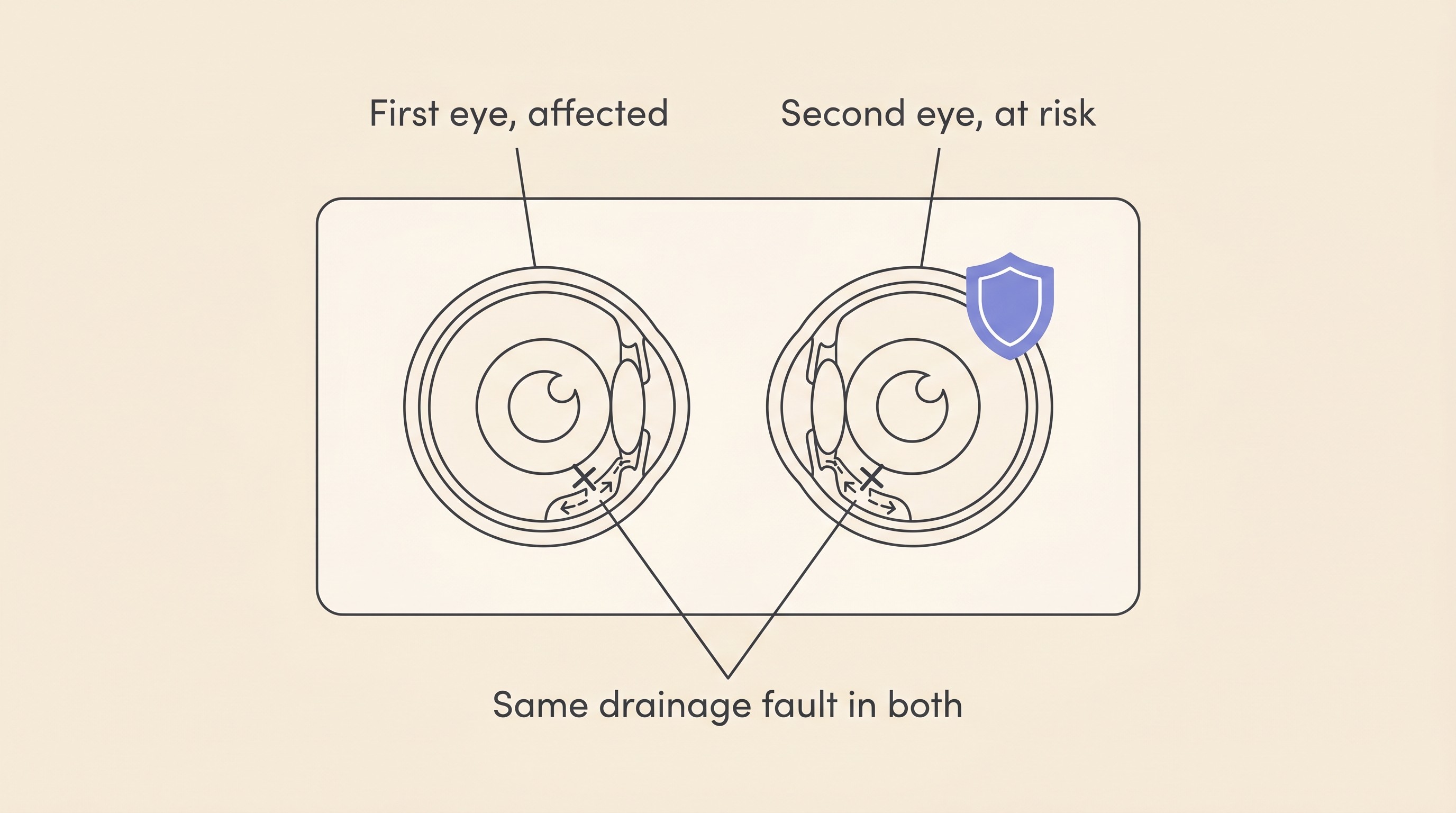
Here's what makes primary glaucoma different from most eye problems, and it's the key to everything that follows. Primary glaucoma is, at root, a bilateral disease (Reinstein, 2018). The trouble isn't really in the eye that went first. It's in the drainage angle, the tiny mesh at the front of the eye through which fluid is meant to escape, and in primary glaucoma that drainage system carries an inherited fault, present from birth (PDSA). Your dog was born with that fault in both eyes. So when one eye fails, it isn't bad luck striking a single spot, it's a body-wide trait showing its hand, and the eye that's still working has the same faulty plumbing waiting in it. As one UK charity puts it plainly, primary glaucoma "usually starts in one eye and over time usually progresses to both", and "most dogs with primary glaucoma end up losing sight in both their eyes" if nothing is done (PDSA). I don't quote that to frighten you, but because it's exactly why we don't sit on our hands with the second eye.
How real is the risk, in numbers? This is the figure I'd want you to hold. In a dog with acute primary angle-closure glaucoma in one eye, the normal-pressured fellow eye has about a 50% chance of an attack in the other eye within roughly 8 months if nothing is done (Miller, 2008). Roughly a coin toss, within the better part of a year. That's a sobering number, but it's a starting point, not a verdict, because the rest of this is about how much we can shift it.
The genuinely good news: drops buy time
Now the part I most want you to take away: we are not helpless here. Preventive eye drops, started in the good eye, roughly halve that risk and buy time.
The clearest evidence comes from the landmark trial behind the figure. Researchers followed 106 dogs with primary closed-angle glaucoma in one eye. In the untreated dogs the second eye succumbed at a median of about 8 months, while in the dogs given preventive drops that median was pushed out to around 30 to 31 months, a highly significant difference (Miller et al., 2000). In plain terms, the drops shifted the point at which half the eyes had had an attack from about 8 months to about 30 months (Miller, 2008). For many dogs that's the difference between losing the second eye within the year and keeping useful sight for a couple of extra years.
Let me be precise about what that means. The drops buy time, they delay the attack. They are not a cure and not a guarantee, because the underlying disease is progressive and in many dogs prophylaxis postpones the second attack rather than preventing it (Miller et al., 2000). So the honest frame is "we're buying your dog more sighted time, quite possibly years of it", not "we've fixed it".
As for which drops, I'll keep this light, because running the schedule day to day belongs to managing glaucoma at home and the eye-drop and IOP tracker, and your specialist prescribes for your dog. In broad strokes, the agents lower the pressure or cut the fluid the eye makes. The most common modern choices are aqueous-suppressing drops: dorzolamide (a carbonic anhydrase inhibitor) and timolol (a beta-blocker), often combined in one product (Plummer et al., 2021). Betaxolol, the beta-blocker used in the original trial, and historically demecarium bromide with a low-dose night-time steroid, have also been used (Miller et al., 2000; Plummer et al., 2021). The names matter less than the principle: a daily drop in the good eye, started early, on time, for life.
One careful caveat, then I'll hand it to your vet. Latanoprost, a powerful pressure-lowering drop, is used only infrequently as a fellow-eye preventive (Plummer et al., 2021), and there's a good reason behind that caution. It works partly by tightly constricting the pupil, and in an eye with an unstable lens that intense, tight pupil can actually trap a forward-slipping lens and drive the pressure up rather than down, so it's approached carefully, particularly in terrier breeds prone to lens instability. It's a reminder that the obvious strong drop isn't always the right one for the good eye, and exactly why this is your ophthalmologist's call, not a website's.
The honest part: what the newer evidence says
I'd be doing you a disservice if I left it at "drops halve the risk, job done", because the research has moved on and two larger, more recent studies temper that older trial. A 2026 multicentre study of 117 dogs found that within five years, 70.9% of dogs with glaucoma in one eye went on to develop it in the other, at a median time to treatment failure of about 2.15 years (Donohue et al., 2026). So over the long run, most second eyes do eventually follow. That's the sobering truth underneath the hopeful headline, and you're better off hearing it from me now than feeling misled later. That same study found time to failure was no different between one class of preventive drop and another, nor between dogs who did or didn't have a steroid added, concluding "no treatment regimen was found to be superior to another" (Donohue et al., 2026). An earlier study had gently suggested adding an anti-inflammatory drop might help (a median 324 days to failure with one versus 195 without) but openly acknowledged the difference wasn't statistically significant (Dees et al., 2014). The variation is real: a survey of 199 eye specialists found wide differences in which drops they use and concluded there's "a great need for well-designed, prospective, controlled, multi-center studies to determine which protocols have the greatest efficacy" (Plummer et al., 2021).
So what should you take from all that? Not "the drops don't work", and definitely not "it's hopeless". The second eye is at real risk over the years, preventive drops are the standard of care and the best tool we have, and they buy meaningful time even though the perfect recipe is still being worked out. That's a reason to pair them with close watching and fast action, which is the rest of the plan, not a reason to skip them.
The plan: three things that protect the fellow eye
Strip away the science and protecting the good eye comes down to three things you can genuinely do, an active plan rather than anxious waiting.
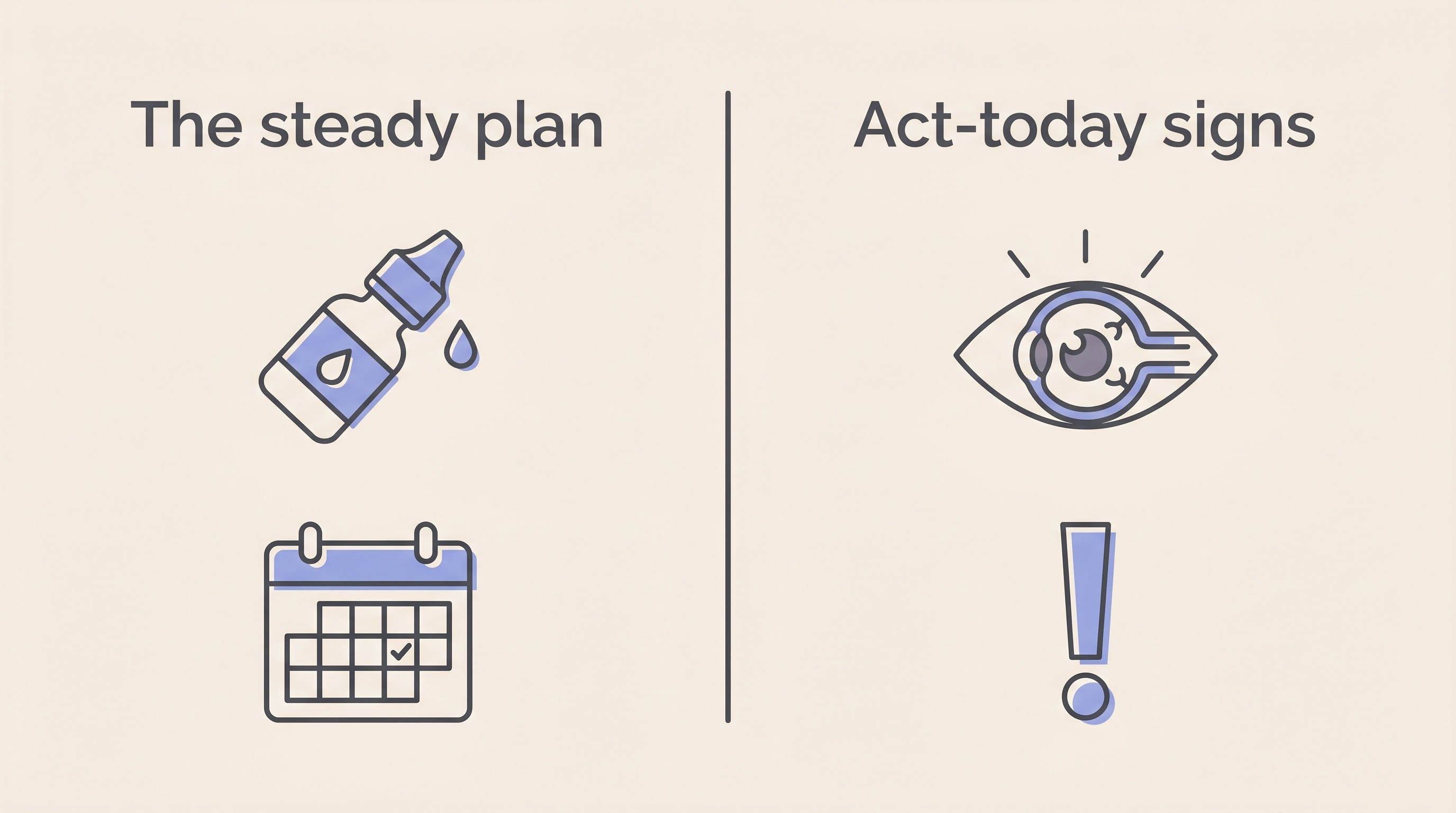
One: the preventive drops, on time, for life. Once your vet recommends them, they aren't optional and they aren't a "when I remember" job. They buy time only if they're actually in the eye, on schedule, every day (Miller, 2008; PDSA). This is the single biggest lever you hold for the second eye. Building the routine, cap-colour cues, reminders, a second person in the early days, is the territory of managing glaucoma at home and the eye-drop tracker, which keeps a per-eye schedule and nudges you when one's due.
Two: regular pressure checks in both eyes. This turns prevention from hope into surveillance. Your vet measures the pressure in the affected and the at-risk eye on a schedule (often every few months once stable), because a pressure that's quietly creeping up can sometimes be caught before it becomes a full, painful, sight-losing attack (Reinstein, 2018; PDSA). For breeds known to carry the inherited drainage fault, pressure screening is sensible even before any diagnosis (Davies Veterinary Specialists). Home pressure monitoring is increasingly possible where your vet supports it, but that detail lives with managing glaucoma at home and the tool's IOP log.
Three: knowing the emergency signs, so you act in hours. This is the one I'd tattoo on the inside of your eyelid if I could. An acute attack in the second eye is a sight-saving emergency measured in hours, not days. If that eye becomes suddenly red, cloudy, painful or blind, it needs a vet today. Here's why the clock is so unforgiving: sustained high pressure kills the retinal ganglion cells, the nerve cells that carry vision to the brain, and in mammals those cells do not regenerate once lost (Komáromy et al., 2019). Vision lost in the first day or two may simply not come back, so speed isn't fussiness, it's the whole game. If you're ever unsure whether what you're seeing counts, the eye-emergency triage sorts it in a minute and the signs are summarised in the eye-emergency red-flags guide. The instinct to "see how it looks in the morning" is the one to override here.
Why this is a primary-glaucoma rule, not a universal one
One important clarification, because I don't want the wrong owner carrying this statistic. Everything above applies to primary glaucoma, the inherited, both-eyes-affected kind. It does not automatically apply to secondary glaucoma, where the pressure rises because of some other problem in that particular eye: chronic inflammation (uveitis), a slipped lens, an advanced cataract, a tumour or an injury (Davies Veterinary Specialists). There's usually no inherited bilateral drainage fault sitting in wait, so the other eye isn't facing the same near-coin-toss. The job there is to find and control the underlying cause, and the second eye's risk depends on whether that cause is one-sided or could affect both eyes too, as a body-wide inflammation might. So if your dog's glaucoma is secondary, the "50% in 8 months" rule isn't yours to carry, and your vet steers the second eye by its actual cause. The depth on the commonest driver, inflammation, lives in uveitis.
A quick word for any cat owner who's wandered in: in cats, glaucoma is almost always secondary, usually to long-standing inflammation inside the eye, so this whole inherited-second-eye story is really a dog one. The feline picture is its own thing, covered in glaucoma in cats.
Where this leaves you and your dog
Let me end where you probably are emotionally, not just clinically. If the first eye has already been removed, that decision may still ache and you may be quietly second-guessing it. Please don't. Taking away a blind, painful eye is one of the kindest operations we do, the relief of constant pain is the entire point of it, and dogs are very often brighter within days. It isn't a failure or a loss, whatever it felt like in the consult room (enucleation and eye removal is written for exactly this moment). And if the second eye is lost too one day, a fully blind dog can still live a genuinely happy, full life, leaning on a remarkable nose and ears, which is what the living-with-vision-loss guides are for. I'd never want you facing the future thinking blindness means a poor life, because it really doesn't.
But that's the worst case, and your job now is to make it less likely. The three-part plan above is the most any of us can do, and it genuinely buys time, quite possibly years of it. So set up the eye-drop and IOP tracker today so the routine runs itself, keep the eye-emergency triage bookmarked where you can find it at 2am, and book that next pressure check before you leave this page. The first eye was the warning. The second eye is the one you get to fight for, and you're already doing it.
References
- Davies Veterinary Specialists. (n.d.). Glaucoma Fact Sheet.
- Dees, D. D., Fritz, K. J., MacLaren, N. E., Esson, D. W., Sheehan Gaerig, A. M., Atkins, R. M., & Knollinger, A. M. (2014). Efficacy of prophylactic antiglaucoma and anti-inflammatory medications in canine primary angle-closure glaucoma: a multicenter retrospective study (2004-2012). Veterinary Ophthalmology, 17(3), 195-200.
- Donohue, L. K., Bentley, E., Boush, J. J., Lasarev, M. R., Pumphrey, S. A., Maggio, F., & Yang, V. Y. (2026). Factors Influencing the Incidence and Onset of Primary Angle-Closure Glaucoma in the Unaffected Eye of Dogs. Veterinary Ophthalmology, 29(1), e70038.
- Komáromy, A. M., Bras, D., Esson, D. W., Fellman, R. L., Grozdanic, S. D., Kagemann, L., Miller, P. E., Moroi, S. E., Plummer, C. E., Sapienza, J. S., Storey, E. S., Teixeira, L. B., Toris, C. B., & Webb, T. R. (2019). The future of canine glaucoma therapy. Veterinary Ophthalmology, 22(5), 726-740.
- Miller, P. E. (2008). Acute Primary Angle-Closure Glaucoma. Clinician's Brief.
- Miller, P. E., Schmidt, G. M., Vainisi, S. J., Swanson, J. F., & Herrmann, M. K. (2000). The efficacy of topical prophylactic antiglaucoma therapy in primary closed angle glaucoma in dogs: a multicenter clinical trial. Journal of the American Animal Hospital Association, 36(5), 431-438.
- PDSA (People's Dispensary for Sick Animals). (n.d.). Glaucoma in dogs. PDSA Pet Health Hub.
- Plummer, C. E., Bras, D., Grozdanic, S., Komáromy, A. M., McLellan, G. J., Miller, P. E., Sapienza, J. S., Teixeira, L., & Webb, T. (2021). Prophylactic anti-glaucoma therapy in dogs with primary glaucoma: A practitioner survey of current medical protocols. Veterinary Ophthalmology, 24(Suppl 1), 96-108.
- Reinstein, S. (2018). Acute Glaucoma: A True Emergency. Today's Veterinary Practice, 8(2), 38-46.
Keep track of how your pet is doing
The owners who cope best are the ones who notice changes early. A simple health log shows you what is working, and what is not, before the next vet visit.
Start tracking, freeYou're not doing this alone
Compare treatment journeys and talk to owners managing vision & eye health. Free to join.
Join PetsLikeMine