Cataracts in Dogs and Cats: What They Are and Why They Form
Dr. Alastair Greenway
MRCVS
When I tell an owner their dog has a cataract, I can usually see two things happen at once. There's the worry, of course, the word carries a lot of weight. But there's often relief too, because they'd been peering into that cloudy eye for weeks fearing the worst, and now at least it has a name. So before we get anywhere near decisions, let me do what I'd do in the consult room: take the word apart and explain, in plain English, what a cataract actually is, why it forms, and what it does and doesn't mean for your pet.
The first thing to say is that a cataract is not the same as the gentle bluish haze a lot of older dogs and cats develop. That haze is usually nuclear sclerosis (also called lenticular sclerosis), a normal ageing change of the lens that tends to be noticed after eight to ten years of age and still lets light through, so the pet sees fine (Conway, 2023; Cornell Riney, n.d.). A cataract is different: it genuinely blocks light. The catch is that the two look very similar from the outside, and even we can't tell them apart reliably without dilating the pupil and shining a light in. If "is this just age?" is really your question, that whole reassurance is covered in is my pet's cloudy eye something to worry about. Here, I'll take it your vet has looked and called it a cataract, and explain what that means.
What a cataract actually is
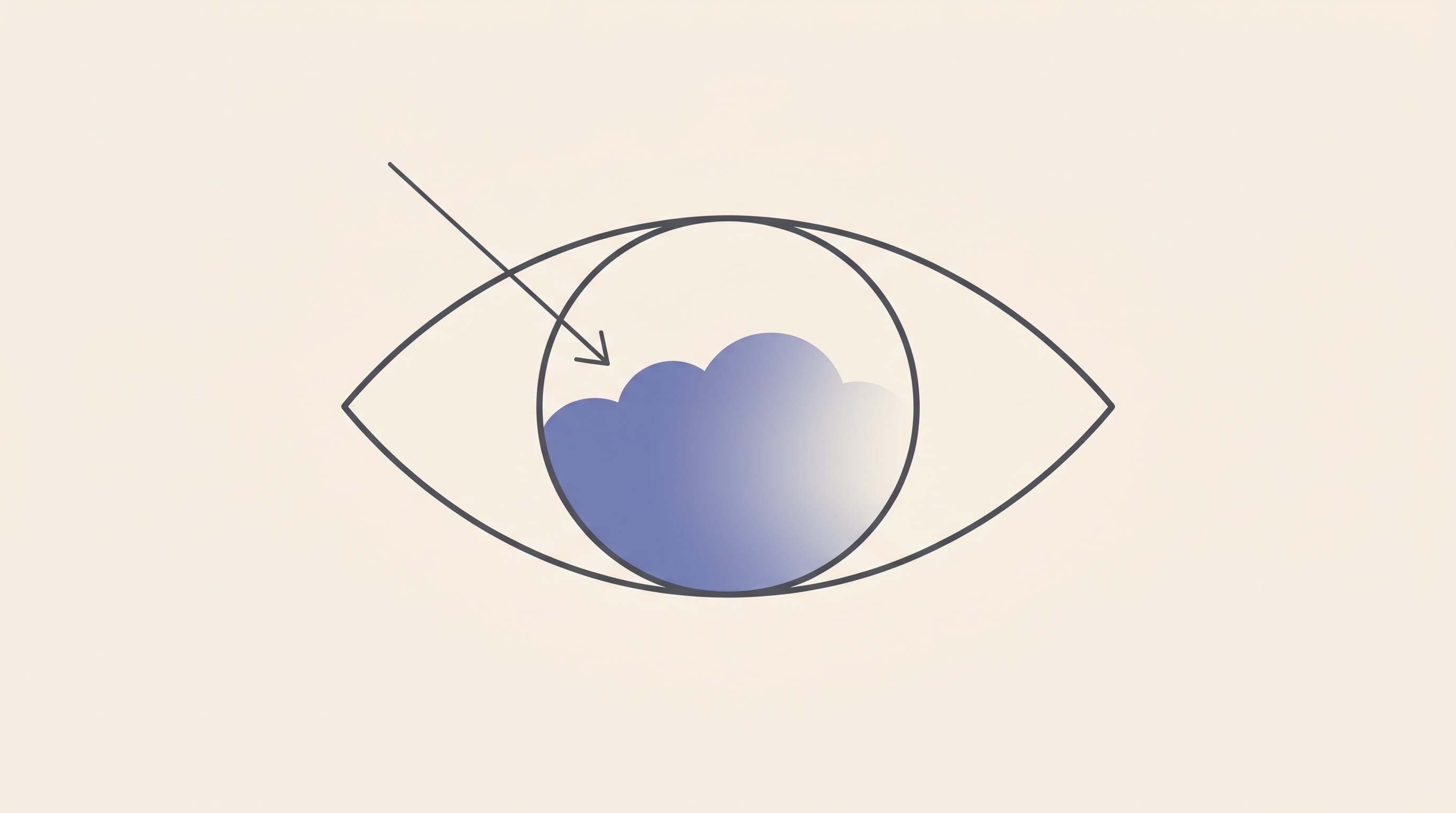
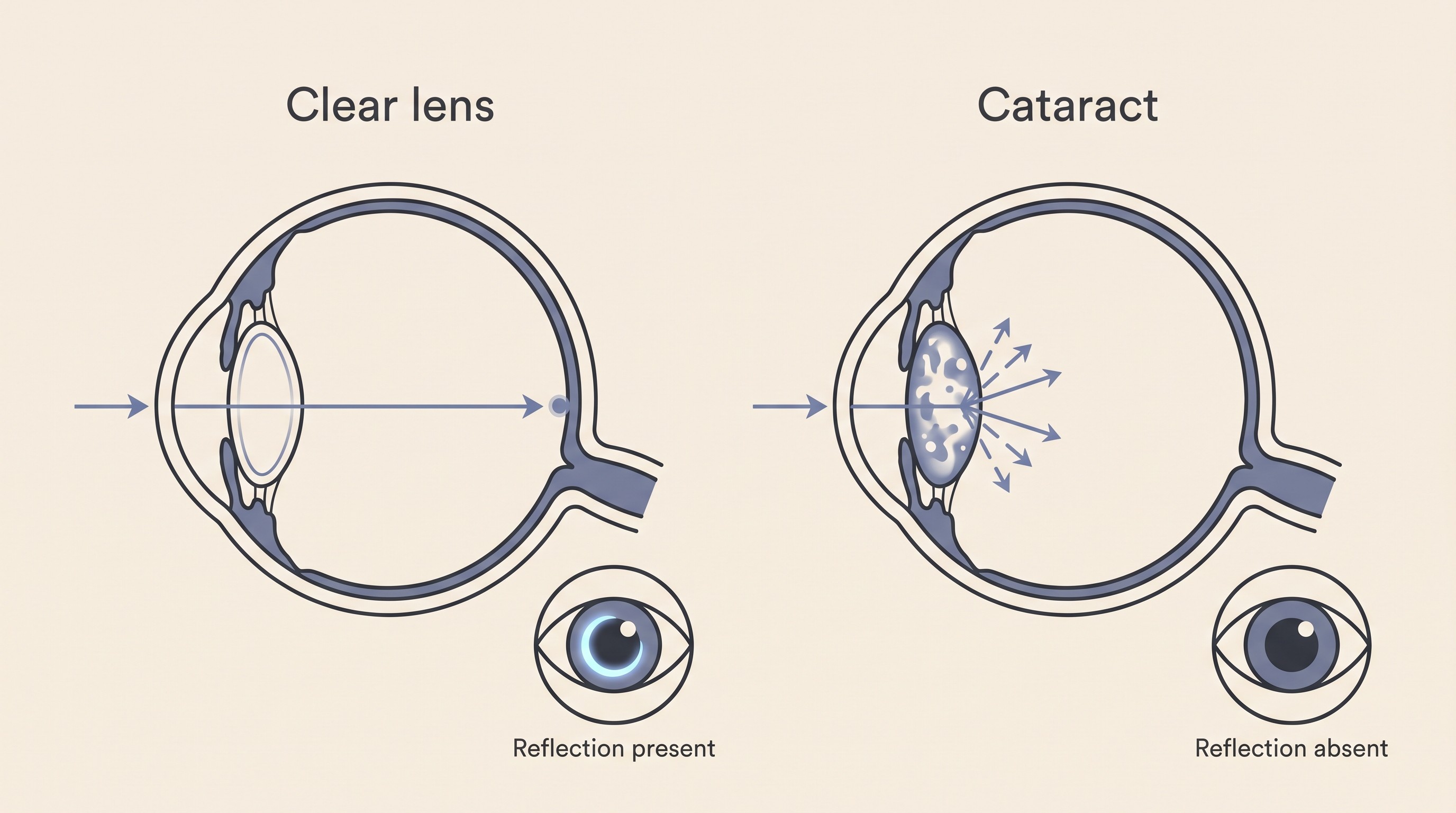
Just behind the pupil sits the lens: a small, normally crystal-clear disc whose job is to focus incoming light onto the retina at the back. It stays that clear through an extraordinarily ordered internal structure, its proteins arranged so precisely that light passes straight through as if through glass. A cataract is what happens when that order breaks down. The proteins clump together, the tissue goes from clear to cloudy, and light can no longer pass cleanly to the retina (Conway, 2023; Cornell Riney, n.d.). If only part of the lens is affected, vision in that eye is blurred or patchy. If the whole lens clouds over, that eye is effectively blind, because the picture can't get through to be sent on to the brain (Cornell Riney, n.d.). The lens has stopped being a clear window and become a frosted one.
Why it happened, and why it isn't your fault
This is the question most owners really want answered, and it usually carries a thread of guilt I'm keen to cut. In the great majority of cases the cause is something written into your dog long before you met them, or a metabolic process you couldn't have prevented. Here are the main routes a cataract forms.
Inherited (genetic) cataract. In dogs, this is the single most common cause. Inherited cataracts turn up across a long list of breeds and can appear in young, juvenile or adult dogs (Cornell Riney, n.d.; Fischer, 2019). If your dog's cataract has shown up without diabetes and without an injury, genetics is the likeliest explanation, and that's nothing you did. Which breeds carry which inherited eye problems sits in the round-up of inherited eye conditions.
Diabetic cataract. This is the leading metabolic cause in dogs, and it behaves differently from the others. Most diabetic dogs develop cataracts, around 80% within the first year or so, whatever the glucose control, because the trigger is sugar getting into the lens rather than how well the diabetes is managed (Beam et al., 1999; Cornell Riney, n.d.). They can also form fast, often within five to six months of the diabetes starting, sometimes clouding a lens over in a matter of days to weeks (Beam et al., 1999; Kador et al., 2010). That speed, and the guilt that comes with it, deserves its own piece, so the full eye-angle, including why early referral matters, lives in diabetic cataracts in dogs, and the glucose-control side belongs over in the Diabetes space. The figure to carry away is that it's common and dog-specific, and not a sign you failed your dog.
Age-related (senile) cataract. Separate from the harmless sclerosis haze, genuine cataracts can also form simply with age as the lens proteins gradually change over the years, and these tend to come on slowly (Fischer, 2019).
Secondary cataract. Sometimes the cataract is a consequence of another problem inside the eye, most often chronic inflammation (uveitis), but also glaucoma or progressive retinal atrophy (PRA) (Fischer, 2019; Cornell Riney, n.d.). It runs both ways: a cataract can cause inflammation and inflammation can cause a cataract, which is why your vet looks at the whole eye rather than just the cloudy lens.
Traumatic cataract. A direct blow, or a penetrating injury such as a cat scratch or a thorn, can damage the lens and trigger a cataract (Fischer, 2019; Cornell Riney, n.d.).
Congenital and nutritional cataract. Less commonly, a pet is born with a cataract, sometimes following an infection before birth, and orphan puppies hand-reared on an unsuitable milk replacer can develop nutritional cataracts (Cornell Riney, n.d.). These less usual forms are also the ones most likely to stay stable or even partly clear, which I'll come back to.
So when an owner asks "why mine, what did I do?", the honest answer is almost always: nothing. In dogs the cause is usually genes, age or diabetes, none of which you control. That reassurance is real, and I give it freely. But I won't leave it there, because a cataract is more than a cosmetic cloud, and the reason it needs watching comes a little further down.
A quick word on cats
Cataracts are very much a dog story, and cats need only a short paragraph here. They're uncommon in cats, and when a cat does develop one it's usually secondary to long-standing inflammation inside the eye rather than inherited or diabetic, so a feline cataract sends us hunting for an underlying cause rather than treating the lens in isolation (Cornell Riney, n.d.). Cats are also largely spared the diabetic cataracts that hit diabetic dogs so reliably. The reason is a neat piece of biochemistry: the diabetic mechanism runs through an enzyme called aldose reductase, which converts the excess sugar flooding the lens into sorbitol, a compound that draws water in, swells the lens fibres and clouds the lens (Kador et al., 2010). Dogs have high lens aldose-reductase activity, and older cats, the ones who actually tend to get diabetes, have very little, so the same flood of sugar doesn't do the same damage (Richter et al., 2002; Kador et al., 2010). The feline picture in full, including the infections worth ruling out, is owned by cataracts in cats.
How a cataract progresses
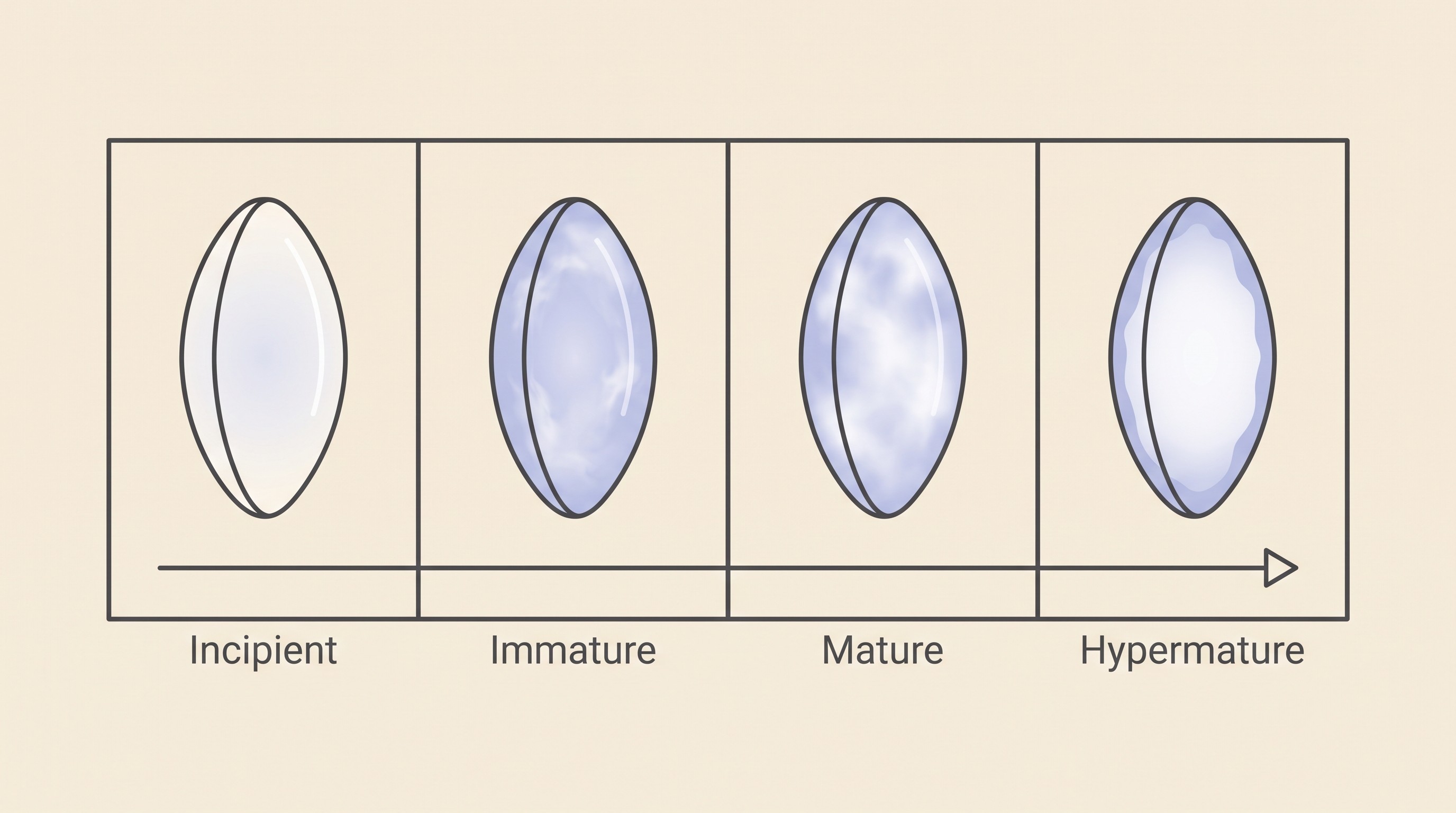
Cataracts are graded by how much of the lens is involved, and that grade is genuinely useful, because it predicts both how much vision is affected and how an eye is likely to do if surgery is ever considered (Fischer, 2019; Conway, 2023). There are four stages.
Incipient. The earliest stage, affecting only about 10 to 15% of the lens, with no obvious effect on vision (Fischer, 2019; Conway, 2023). You might not notice anything wrong at all.
Immature. Now more of the lens is involved, anywhere from around 15% up towards complete. Light still gets partly through, so on examination we can still see some of the reflection off the back of the eye, and vision is variably reduced, from barely affected to nearly blind in that eye depending on how dense the cataract is (Fischer, 2019).
Mature. The whole lens is now cloudy. No reflection from the back of the eye gets through and the eye is functionally blind on that side, though it usually still reacts to a bright light and the pupil still responds, which is one reason your vet checks more than just whether the lens looks cloudy (Fischer, 2019; Cornell Riney, n.d.).
Hypermature. Here the lens begins to break down and resorb. The capsule wrinkles and develops white plaques and shiny sparkles, and inflammation inside the eye is common at this stage. Occasionally enough of the lens clears that a little vision returns, sometimes called a resorbing or Morgagnian cataract, but this is unpredictable and is not a cure (Fischer, 2019; Cornell Riney, n.d.).
This progression isn't a fixed conveyor belt, though. Some cataracts, especially the congenital and nutritional ones, can stabilise or even partly resorb. Diabetic cataracts tend to march quickly. Many inherited cataracts progress over months to years (Fischer & Meyer-Lindenberg, 2018). So a diagnosis of "cataract" doesn't tell you how fast, and watching how your pet's actual sight changes over time is more useful than any single label. The at-home vision check is built for exactly that: a simple, repeatable way to track your pet's functional vision so you see a real trend rather than guess.
Why a cataract is more than a cosmetic cloud
This is the part I most want owners to take away, because it's where the genuinely useful action lives. As a cataract matures, the lens leaks proteins, and the immune system, which normally never meets these proteins up close, reacts to them. That reaction is an inflammation inside the eye called lens-induced uveitis (Cornell Riney, n.d.; Fischer, 2019). It's the most common complication of an untreated cataract and the engine behind most of the trouble cataracts cause: in one referral series it was present in around 71% of eyes assessed before cataract surgery (Fischer, 2019).
Left unchecked, that inflammation can drive two more serious problems. The first is secondary glaucoma, a painful and sight-threatening rise in pressure inside the eye. Lens-induced uveitis is in fact the most common cause of secondary glaucoma in dogs, behind around 81% of cases, and roughly 20% of dogs with cataracts are reported to develop glaucoma at some point (Fischer, 2019). The second is lens luxation, where the lens slips out of position, more likely with long-standing hypermature cataracts and reported in around 6% of cases (Fischer, 2019). Retinal detachment is a further, less common possibility (Fischer, 2019; Cornell Riney, n.d.). Looking across all stages, one study of 447 affected eyes found complications in around 44% of cataracts, more often the more advanced the cataract, with lens-induced uveitis the commonest (Fischer & Meyer-Lindenberg, 2018).
I'm not telling you this to frighten you. Most of these complications are manageable, and many cataracts cause no immediate drama at all. I'm telling you because it's the reason a cataract earns ongoing attention even if you decide against surgery. A cloudy lens itself isn't usually painful, but the inflammation and pressure rises it can set off genuinely are, and they're treatable, especially if caught early. That's why even a non-operated cataract should have periodic eye checks, often every four to six months once it is advanced, and why a cataractous eye that suddenly turns red, painful or visibly worse is a reason to ring your vet rather than wait (Cornell Riney, n.d.). The glaucoma here is owned by glaucoma in dogs, the inflammation by uveitis.
The two roads from here
Once you understand what a cataract is, the choices ahead stop feeling arbitrary. There are, honestly, two.
The first is surgery, the only way to restore vision in a cataractous eye, because no eye drop, supplement or medicine clears a cataract, whatever the internet promises. The good news is that it works well: in dogs, phacoemulsification (breaking up and removing the cloudy lens with ultrasound, usually replacing it with an artificial lens) restores useful vision in the large majority of eyes, with success commonly reported in the region of 80 to 90%, and it does best on recent cataracts in otherwise healthy eyes (Cornell Riney, n.d.; Krishnan et al., 2020). Whether it's right for your pet is a real decision, with cost, candidacy and timing all in play, and that whole conversation is owned by the cataract surgery decision, with the operation and recovery itself in cataract surgery and recovery.
The second road is to manage and monitor. Choosing not to operate is a perfectly legitimate decision, not a second-best one. Many pets live well with cataracts, especially slowly progressing ones, supported by anti-inflammatory drops to keep that lens-induced uveitis in check and regular pressure checks to catch glaucoma early (Cornell Riney, n.d.; Fischer, 2019). There's an honest nuance here worth knowing. When researchers compared operated and non-operated eyes, the eyes their ophthalmologist had judged poor surgical candidates ran into far more trouble than the eyes whose owners had simply chosen against an operation on an otherwise healthy eye, so an eye that's a good candidate often does well whichever path you take, while a problem eye needs watching closely whatever you decide (Krishnan et al., 2020). How to do that watchful management properly is covered in living with cataracts without surgery.
So here's where I'd leave you. A cataract means the lens has gone cloudy and is blocking light, and the why is almost always genes, age or, in a diabetic dog, the diabetes, not anything you did. It isn't usually an emergency, and a pet can live a good life with one, especially a slowly progressing one, but it's more than cosmetic because of the painful complications it can quietly cause. So the useful next steps are small and concrete: find out which kind of cataract you're dealing with, watch for any sudden redness, pain or worsening and act if it comes, track your pet's real-world sight over time, and then decide, unhurried, between surgery and careful monitoring. If you'd like to start weighing that last choice, the cataract surgery decision is the place to go next.
References
- Beam, S., Correa, M. T., & Davidson, M. G. (1999). A retrospective-cohort study on the development of cataracts in dogs with diabetes mellitus: 200 cases. Veterinary Ophthalmology, 2(3), 169-172.
- Conway, E. (2023, April 14). Cataracts versus nuclear sclerosis. American College of Veterinary Ophthalmologists (ACVO Public).
- Cornell University College of Veterinary Medicine, Riney Canine Health Center. (n.d.). Canine cataracts.
- Fischer, M. C. (2019). Canine cataracts: classification, aetiology and complications. Vet Times.
- Fischer, M. C., & Meyer-Lindenberg, A. (2018). Progression and complications of canine cataracts for different stages of development and aetiologies. Journal of Small Animal Practice, 59(10), 616-624.
- Kador, P. F., Webb, T. R., Bras, I., Ketring, K., & Wyman, M. (2010). Topical Kinostat ameliorates the clinical development and progression of cataracts in dogs with diabetes mellitus. Veterinary Ophthalmology, 13(6), 363-368.
- Krishnan, H., Hetzel, S., McLellan, G. J., & Bentley, E. (2020). Comparison of outcomes in cataractous eyes of dogs undergoing phacoemulsification versus eyes not undergoing surgery. Veterinary Ophthalmology, 23(2), 286-291.
- Richter, M., Guscetti, F., & Spiess, B. (2002). Aldose reductase activity and glucose-related opacities in incubated lenses from dogs and cats. American Journal of Veterinary Research, 63(11), 1591-1597.
Keep track of how your pet is doing
The owners who cope best are the ones who notice changes early. A simple health log shows you what is working, and what is not, before the next vet visit.
Start tracking, freeYou're not doing this alone
Compare treatment journeys and talk to owners managing vision & eye health. Free to join.
Join PetsLikeMine