Diabetic Cataracts: Why Most Diabetic Dogs Go Cloudy, and Why Speed Matters
Dr. Alastair Greenway
MRCVS
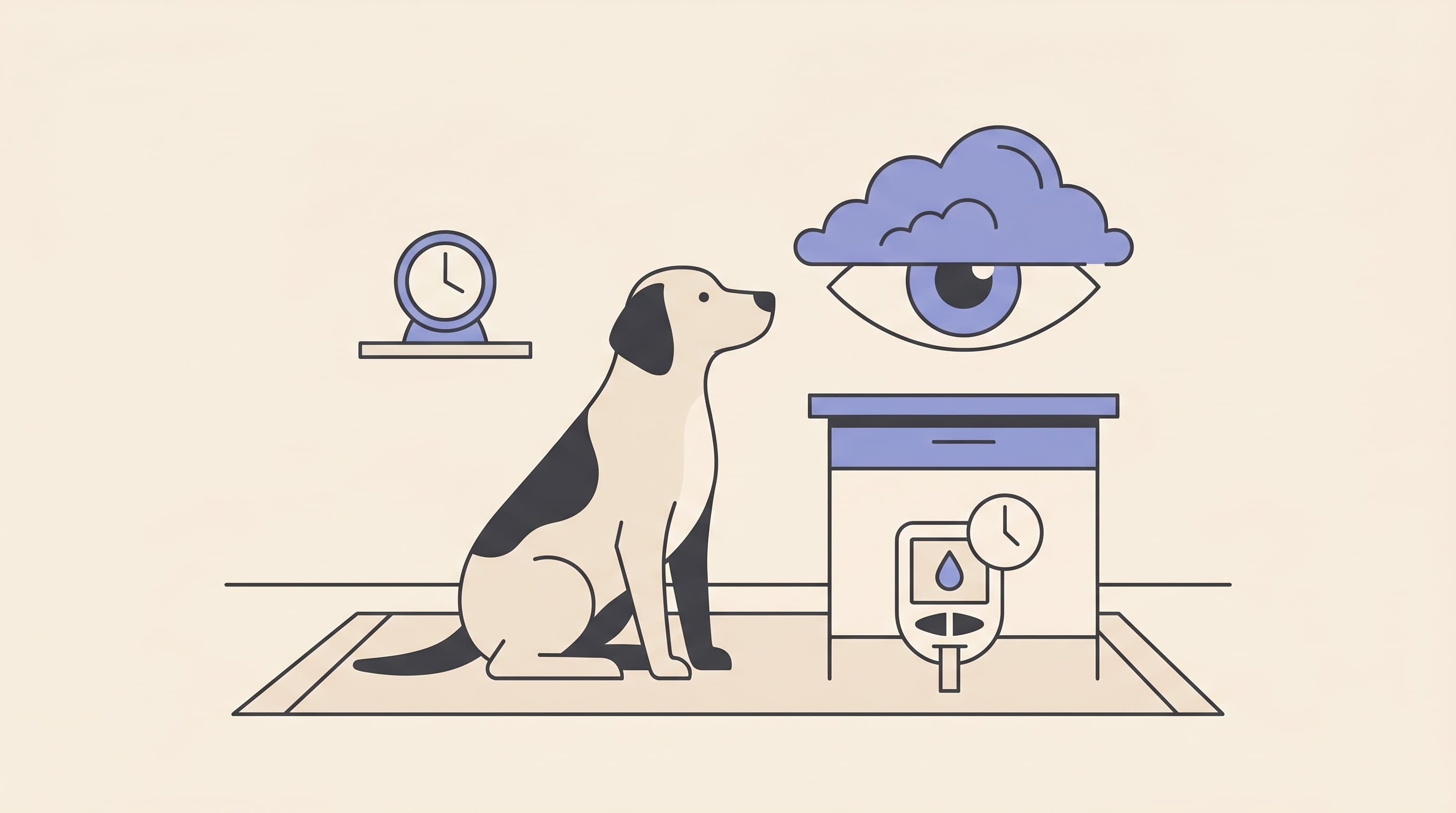
There's a particular phone call I've taken many times over the years, and it almost always opens the same way. A dog was diagnosed with diabetes a few months ago, the owner has worked hard at the injections and the feeding times, and now the eyes are going cloudy. Underneath the worry, nearly every time, is a quiet, awful question: did I do this?
So before anything else, let me say the thing I most want you to hear. You almost certainly did not cause this, and no realistic change in your injections would have prevented it. Cataracts are simply what diabetes does to a dog's eyes. Most diabetic dogs get them, around 80% within the first year or so, regardless of how carefully the diabetes is managed. That isn't me being kind, it's what the evidence says. Once that weight is off you, we can talk clearly about what actually matters now: why a clouding diabetic eye needs seeing promptly rather than watching, and why getting in early gives your dog the best chance of seeing again.
Most diabetic dogs get cataracts, and it isn't your fault
Let me give you the number, because the number is what relieves the guilt. In the landmark study that the whole profession still quotes, researchers followed 177 diabetic dogs who had clear lenses at the time of their diabetes diagnosis. Of those, 132 went on to develop cataracts. Half had them by around five to six months after diagnosis, three-quarters by about a year, and roughly 80% within around sixteen months (Beam et al., 1999). Cornell put the same finding in plain owner language: about 75 to 80% of diabetic dogs develop cataracts within the first year of diagnosis, "regardless of how well-controlled their diabetes is" (Cornell Riney, n.d.). And in a ten-year referral series, cataract turned out to be far and away the commonest eye finding in diabetic dogs, present in 97.3% of the affected eyes that came through the door (Cantero et al., 2023).
So if your diabetic dog's eyes are clouding, you are in the large majority, not some unlucky minority who "must have done something". This is expected, it's part of the disease, and the useful question is never "what did I get wrong", it's "what do we do now".
Why it happens: it's the sugar in the lens, not the dog's age
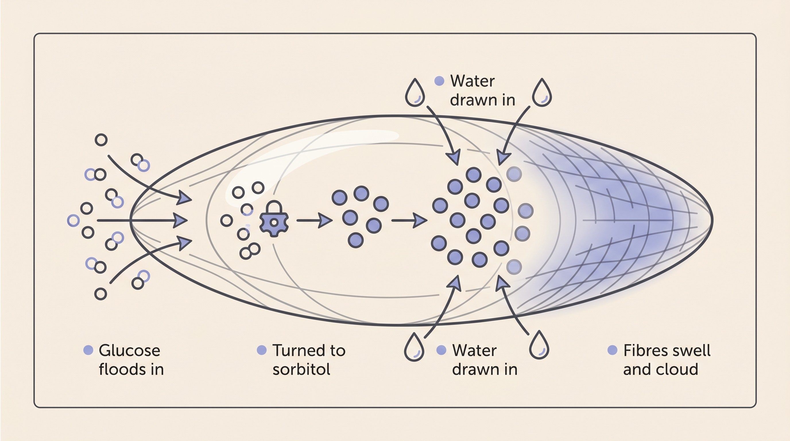
The mechanism is worth a moment, because it explains both why it isn't your fault and why it can come on so fast. The lens, the clear disc behind the pupil that focuses light, stays crystal clear through an extraordinarily ordered internal structure. When blood glucose runs high, glucose floods into the lens, the enzyme that usually handles it gets overwhelmed, and the excess is shunted down a side route called the polyol pathway, where an enzyme named aldose reductase converts it into a compound called sorbitol (Lee & Chung, 1999; Cornell Riney, n.d.). Sorbitol is the troublemaker: it draws water in and gets trapped inside the lens fibres, so water follows, the fibres swell and rupture, the structure breaks down, and the lens turns opaque, with oxidative stress compounding the damage (Lee & Chung, 1999; Richter et al., 2002; Cornell Riney, n.d.).
The takeaway from all that biochemistry is simple. This is driven by sugar getting into the lens, not by your dog getting older and not by anything you did with the needle. It's also why it can happen quickly, and why a diabetic cataract behaves so differently from the slow, age-related cloudiness other dogs get. (For the full anatomy of what a cataract is and the other ways they form, see cataracts in dogs and cats; here I'm staying with the diabetic story.)
One more thing this mechanism explains: why cats are largely spared. The whole cascade runs on aldose reductase, and the lenses of older cats have markedly lower activity of it than the lenses of dogs or young cats. Because feline diabetes typically starts after about seven years of age, the enzyme that drives the damage is largely quiet by the time a cat becomes diabetic, so cats rarely develop diabetic cataracts despite identical high blood sugar (Richter et al., 2002). The feline cataract picture, which is a different story usually rooted in inflammation rather than sugar, lives in cataracts in cats.
Why speed matters, part one: they can blind a dog fast
Here the message shifts from reassurance to action. Diabetic cataracts often come on quickly: where an inherited or age-related cataract might creep over months or years, a diabetic one can go from a clear lens to a blinding one over days to a few weeks, and it usually affects both eyes at once (Cornell Riney, n.d.). Owners often describe a dog that seemed fine, then within a fortnight was hesitating on the stairs, missing a thrown treat, or bumping into furniture that hadn't moved. Because both eyes tend to cloud together, there isn't a good eye to lean on, so the change in how the dog gets about can feel like it arrives overnight. In one case series of diabetic dogs that came to referral, the cataracts had been present on average only about five to six weeks before surgery (Wilkie et al., 2006).
So with diabetes, "watch and wait" is the wrong setting. The cloudiness you noticed this week may be markedly worse next week. That isn't a reason to panic, but it is a reason not to sit on it.
One quick distinction, then I'll hand off. The faint bluish-grey haze many older dogs get is usually nuclear sclerosis, a normal ageing change that doesn't blind, whereas a diabetic cataract is a genuine, light-blocking opacity of the lens. The honest catch is that you can't reliably tell them apart by looking, and neither can I without examining the eye (Cornell Riney, n.d.). But in a diabetic dog the index of suspicion is already high, so a newly cloudy eye earns a prompt look, not a "let's see how it goes". The full cloudy-eye sorting is owned by is my pet's cloudy eye something to worry about.
Why speed matters, part two: the swelling lens can actually hurt
Here's the part owners rarely know, and it's why a diabetic cataract is more than a cosmetic cloud. As the lens takes on water it doesn't just cloud, it swells: a normal canine lens of roughly 7mm can swell towards 10mm, and that enlargement carries real risk (NDSR, n.d.).
A swollen, leaky lens lets lens proteins escape, and the immune system, which normally never meets these proteins, reacts to them. That reaction is an inflammation inside the eye called lens-induced uveitis, and it's painful (NDSR, n.d.; Cornell Riney, n.d.). In the worst cases the lens swells so dramatically that its capsule, the thin bag that holds it, ruptures of its own accord, spilling lens material into the eye and triggering severe inflammation that risks secondary glaucoma and even retinal detachment (Wilkie et al., 2006; Eastcott Referrals, n.d.). This isn't rare in the dogs who reach referral: in one diabetic series the great majority of cataractous eyes had a spontaneous lens-capsule rupture before surgery, and the swollen, intumescent type was the single commonest pattern in another large series (Wilkie et al., 2006; Cantero et al., 2023).
The signs to watch for are a red, sore or squinting eye, a dog rubbing at its face, or an eye that suddenly looks worse. Any of those in a diabetic dog deserves prompt veterinary attention, because it can mean the lens is inflaming the eye (Cornell Riney, n.d.). It's also why even a diabetic cataract you decide not to operate on still needs anti-inflammatory eye drops and monitoring: the cataract itself may be painless, but the inflammation it sets off is not, and that part is treatable (Eastcott Referrals, n.d.).
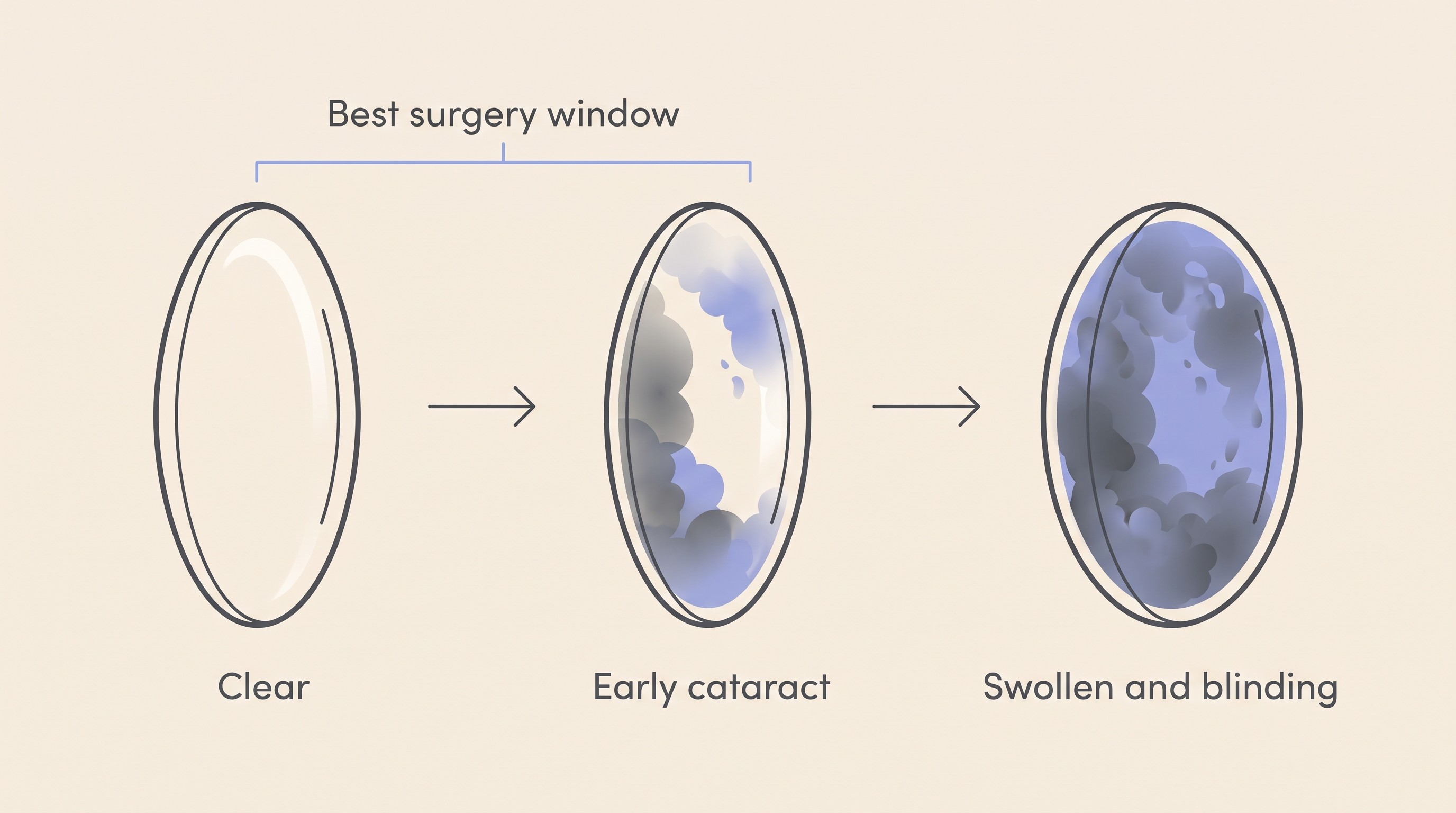
Why speed matters, part three: surgery works best on a recent cataract
Now the genuinely hopeful part, and the reason early really does beat waiting. The only treatment that restores vision in a cataractous eye is surgery, specifically phacoemulsification, where the cloudy lens is broken up and removed by ultrasound and usually replaced with an artificial one (ACVO, n.d.). No drop, supplement or diet clears a cataract, whatever the internet promises. And it works genuinely well in diabetic dogs. In a direct comparison, 94.8% of diabetic eyes treated with phacoemulsification were visual at the final check, against just 7.6% of eyes managed with drops alone, and the drops-only eyes carried nearly four times the risk of complications (Lee et al., 2023). For cataract surgery in dogs more broadly, the reported success in ideal candidates is high, in the order of 80 to 90% of eyes regaining functional vision, with the best outcomes when the surgery is done sooner rather than later (Cornell Riney, n.d.). So the honest headline is that most operated eyes regain useful sight, and the diabetic numbers sit right in that range. The realistic figure for your individual dog depends on the state of the eye, which is exactly what the referral assessment is for, and not every eye will be a candidate.
But timing matters. Advanced, long-standing cataracts are, in Cornell's words, "more likely to cause lens-induced intraocular inflammation, lens instability, and loss of lens capsule integrity", all of which can worsen the outcome or even take surgery off the table (Cornell Riney, n.d.). A recent cataract in an otherwise healthy eye is a far better surgical prospect than one that's been swelling and inflaming for months. UK ophthalmologists therefore tend to advise operating on a mature diabetic cataract promptly, often within a couple of weeks of it being spotted, and they warn specifically against waiting for "perfect" diabetic stabilisation before referring, because that wait can quietly cost the eye (NDSR, n.d.; Eastcott Referrals, n.d.). It is, as one UK referral centre puts it, a race against time (Eastcott Referrals, n.d.). Early referral keeps the eye in the window where surgery does best, which is the heart of why a clouding diabetic eye is a prompt-referral situation, not a wait-and-see one.
What to do now, in order
If your diabetic dog's eyes are starting to cloud, here's the practical sequence.
Tell your vet promptly and ask them to examine the eyes. Don't wait for the next routine diabetes check, and treat a red, sore or suddenly-worse eye as a reason to ring the same day.
Ask about referral to a veterinary ophthalmologist. This is the conversation that decides whether surgery is on the cards, and earlier is better. Not every diabetic dog is a surgical candidate, and finding out is part of the point. A specialist checks that the retina behind the cloudy lens still works, with an electroretinogram, and rules out a detached retina hiding behind the opacity, with an ocular ultrasound, because a cataract can mask retinal disease, and operating on an eye whose retina has already failed would restore a clear lens but no sight (Cornell Riney, n.d.; Lee et al., 2023). A reasonably stable, anaesthesia-fit diabetic is also part of being a candidate, which is one reason the referral conversation is worth having alongside your vet rather than instead of them. The full operate-or-not decision, the candidacy criteria, the cost and the legitimate no-surgery path are owned by the cataract surgery decision, and the things worth asking at that consult are gathered in questions to ask before cataract surgery (there is a printable worksheet at cataract surgery questions).
Ask about anti-inflammatory eye drops in the meantime. Because the swelling lens can inflame the eye, UK ophthalmologists advise starting anti-inflammatory treatment as soon as a diabetic cataract is noted, whether or not surgery is planned, to keep that lens-induced uveitis in check (Eastcott Referrals, n.d.).
Keep tracking your dog's actual vision. A label tells you less than the trend. Watching how your dog copes week to week, and logging it, gives you and your vet something concrete to act on. Our at-home vision check is built for exactly that.
A word on glucose control, since it's the thing owners most want to influence. Tighter, steadier control is worth doing and may buy some time, but I won't oversell it: cataracts develop "regardless of how well-controlled the diabetes is" (Cornell Riney, n.d.), so good management delays rather than reliably prevents them. The how-to of that control belongs to the Diabetes space and its Glucose Companion tool, not here. Just don't let a cataract that appears despite your best efforts feel like a failure. It isn't one.
What if surgery isn't the path
Sometimes surgery isn't right, whether for cost, candidacy or a dog who wouldn't tolerate weeks of drops and a cone. If that's where you land, it's a legitimate choice, not a defeat. Dogs adapt remarkably well to losing their sight, especially when it comes on over days to weeks rather than instantly, because they navigate far more by a memorised map of their home plus scent and sound than we tend to credit. A blind but comfortable dog can have a genuinely good life. The work of helping them settle, keeping the furniture where it is, talking before you touch, building safe routines, is real but very doable, and it's owned by your blind dog's first 30 days, with the companion download at first 30 days with a blind pet. The one ongoing job on the no-surgery path is to keep that lens-induced inflammation managed and to act on a red, painful or suddenly worse eye, because comfort matters most.
So here's where I'd leave you, with the next step rather than a platitude. If the eyes are clouding, book the appointment, ask for the cloudy eye to be examined now rather than later, and ask about referral and anti-inflammatory drops while the eye is still recent. That early move is the one most likely to give your dog its sight back. The cloud is common, expected and not your doing, and what happens next is where you make the difference.
References
- ACVO (American College of Veterinary Ophthalmologists). (n.d.). Cataracts. ACVO Public.
- Beam, S., Correa, M. T., & Davidson, M. G. (1999). A retrospective-cohort study on the development of cataracts in dogs with diabetes mellitus: 200 cases. Veterinary Ophthalmology, 2(3), 169-172.
- Cantero, F., Ortillés, Á., Peña, M. T., & Leiva, M. (2023). Prevalence of ocular findings and their association with glycemia in dogs with diabetes mellitus: A 10-year clinical study (2009-2019). Open Veterinary Journal, 13(5), 620-628.
- Cornell University College of Veterinary Medicine, Riney Canine Health Center. (n.d.). Canine cataracts.
- Eastcott Referrals. (n.d.). Avoiding ruptured diabetic cataracts in dogs.
- Lee, A. Y. W., & Chung, S. S. M. (1999). Contributions of polyol pathway to oxidative stress in diabetic cataract. The FASEB Journal, 13(1), 23-30.
- Lee, E., Kang, S., Jeong, D., & Seo, K. (2023). Comparison of the outcomes of phacoemulsification versus topical medication alone in canine diabetic cataracts: a retrospective study. Journal of Veterinary Science, 24(6), e86.
- NDSR (North Downs Specialist Referrals). (n.d.). Diabetic cataracts. Insights.
- Richter, M., Guscetti, F., & Spiess, B. (2002). Aldose reductase activity and glucose-related opacities in incubated lenses from dogs and cats. American Journal of Veterinary Research, 63(11), 1591-1597.
- Wilkie, D. A., Gemensky-Metzler, A. J., Colitz, C. M. H., Bras, I. D., Kuonen, V. J., Norris, K. N., & Basham, C. R. (2006). Canine cataracts, diabetes mellitus and spontaneous lens capsule rupture: a retrospective study of 18 dogs. Veterinary Ophthalmology, 9(5), 328-334.
Keep track of how your pet is doing
The owners who cope best are the ones who notice changes early. A simple health log shows you what is working, and what is not, before the next vet visit.
Start tracking, freeYou're not doing this alone
Compare treatment journeys and talk to owners managing vision & eye health. Free to join.
Join PetsLikeMine