My Pet's Eye Looks Cloudy: Should I Worry?
Dr. Alastair Greenway
MRCVS
It usually happens in a particular kind of light. You're looking down at your old dog dozing in the afternoon sun, or your cat is on the windowsill with the light catching just right, and you notice it: a soft bluish-grey haze deep in the eye where there used to be clear black. Your stomach drops, because the word that arrives almost immediately is "cataract", and the word after that is "blindness".
So let me give you the reassuring half first, because most of you need to hear it. In an older pet, a gentle bluish haze in both eyes, in an animal who is otherwise comfortable and still finding their food bowl and the bottom of the stairs, is most often a normal ageing change called nuclear sclerosis, and it barely touches their vision (Cornell Riney Canine Health Center, n.d.; Clode, 2016). It isn't a cataract, it isn't painful, and it doesn't need treating.
But the honest truth has a second half, and it's the reason this article exists. You genuinely cannot tell that benign haze apart from a sight-stealing cataract just by looking, and neither can I, until I've dilated the pupil and shone a light through it. So the real message sits between panic and "it's just old age": usually nothing, occasionally something, and the only way to know which is one proper look.
The common, reassuring cause: nuclear sclerosis
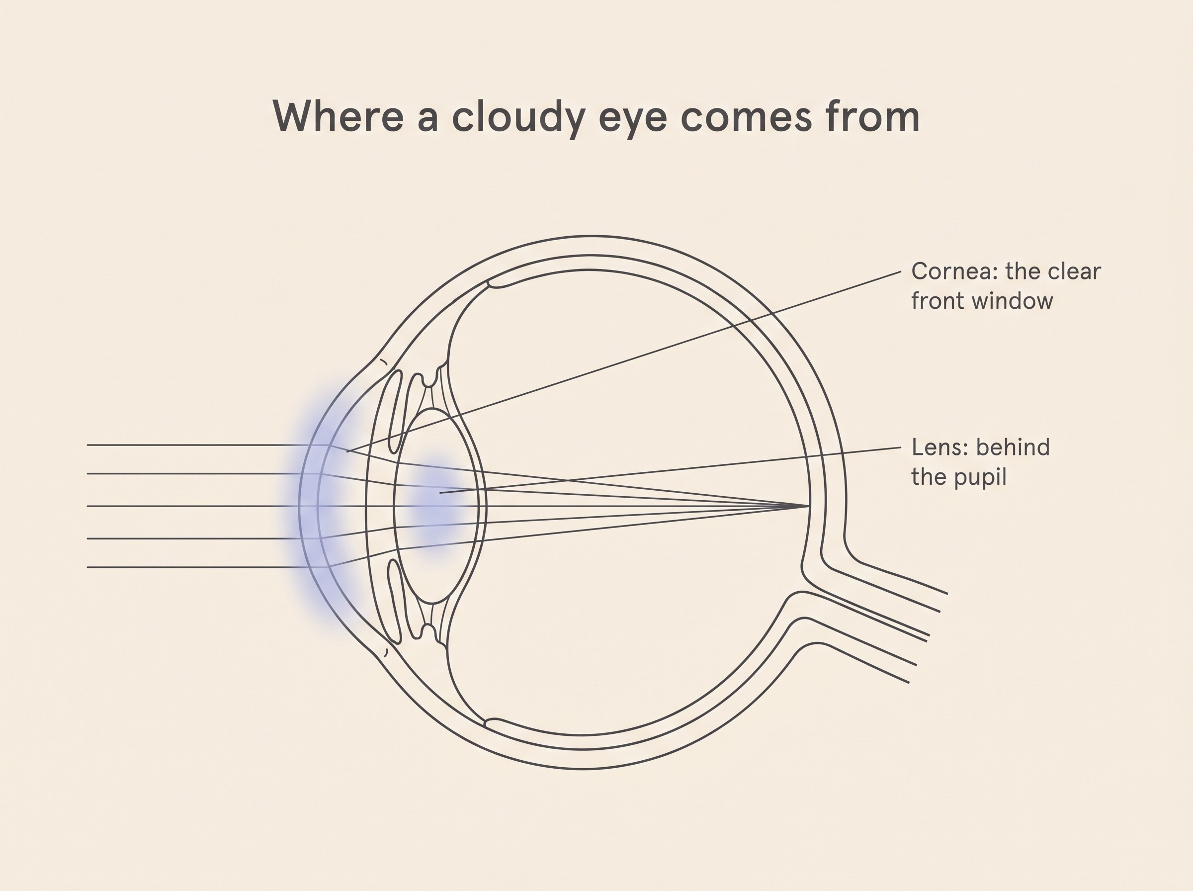
Nuclear sclerosis, sometimes called lenticular sclerosis, is a normal age-related change inside the lens. The lens sits just behind the pupil and keeps laying down new fibres throughout life, like the rings of a tree. Over the years the older fibres in the centre get compressed and a little denser, and that denser core scatters light back as a faint blue-grey cloudiness (Cornell Riney Canine Health Center, n.d.; Clode, 2016). It's very common, turning up in roughly half of dogs over about nine years of age, and it's seen in cats too (Clode, 2016). It's usually in both eyes, fairly evenly, and creeps in so gradually that the day you notice it is rarely the day it started.
Here's the part that should let you breathe out: nuclear sclerosis isn't thought to cause meaningful vision loss (Clode, 2016; Conway, 2023). Light still passes through the lens, it's just travelling through slightly hazier glass. At most an affected pet loses a touch of fine, close-up focus, and honestly that was never their strong suit. A dog or cat sees far less crisp detail than we do and reads the world mostly through nose, ear and movement, so a little extra haze on the fine print costs them almost nothing they were using. It needs no drops and no surgery, and if it's what your vet finds, the appointment ends in reassurance.
The honest catch: you can't tell by looking
Now the bit I won't gloss over, because skating past it is how owners get falsely reassured. A cataract makes the eye look cloudy. So does nuclear sclerosis. To the naked eye the two can look almost identical, whether the eye belongs to you, to a neighbour with strong opinions, or to me (Conway, 2023). So I'll be blunt: nobody can reliably call a cloudy eye "just sclerosis" on appearance alone. It takes an examination.
What does that involve? Your vet puts in a drop to dilate the pupil, usually tropicamide, which widens it like a camera aperture so the whole lens can be seen (Clode, 2016). Then they use a focal light and a technique called retroillumination, which simply means shining light in so it bounces back off the reflective layer at the back of the eye, the tapetum, the layer that makes your pet's eyes flash green in a photo. That returning glow is the clever part. With nuclear sclerosis the glow still comes back, only slightly hazed, because light is still getting through. With a cataract, the opacity interrupts that glow with dark or white patches, because the lens is now blocking the light (Clode, 2016; Conway, 2023). It's quick, painless, and it turns "I'm worried" into a clear answer in minutes.

When the cloudiness really is a cataract
So what is a cataract, the thing everyone fears? It's the lens itself going opaque, any focal or diffuse opacity of what should be a crystal-clear structure (Cornell Riney Canine Health Center, n.d.; Conway, 2023). And unlike sclerosis, a cataract genuinely does block vision, by however much of the lens has clouded. The scale matters: an early cataract clouding only a tenth or so of the lens may cost no useful sight at all, whereas a mature one clouds the whole lens and causes significant impairment (Conway, 2023). It's never purely cosmetic, either: ageing cataracts can leak lens protein and trigger painful inflammation inside the eye, and lead on to secondary glaucoma and lens luxation (Cornell Riney Canine Health Center, n.d.). I'll hand the depth off rather than rebuild it here. What cataracts are, their types and causes, and how they progress all lives in cataracts explained; my job here is just to get you to the right door.
The one cloudy eye you must not wait on: the diabetic dog
There's a single exception to all this calm, and I want a box drawn around it, because it's the one place where "let's keep an eye on it" is the wrong instinct. If your dog is diabetic and the eyes are clouding over, that's an early-referral situation, not a wait-and-see. Around 80% of diabetic dogs develop cataracts within the first year or so of diagnosis, as glucose floods the lens, is converted to a sugar-alcohol called sorbitol, and that draws in water and swells the lens until it clouds (Beam et al., 1999; Cornell Riney Canine Health Center, n.d.). This happens regardless of how good the blood-sugar control is, so please don't read it as something you failed to manage. It's the sugar in the lens, not anything you did.
Speed matters because these cataracts form fast, sometimes turning a clear eye to a blind one in days to weeks, and a rapidly swelling lens is the kind that can rupture its capsule and set off fierce, painful inflammation (Cornell Riney Canine Health Center, n.d.). Surgery also works best on a recent cataract in an otherwise healthy eye, so early contact with your vet protects your dog's options. The full eye-side story is in diabetic cataracts, and the blood-sugar side is covered in our diabetes content. One reassuring species note: this is a dog story. Cats are largely spared diabetic cataracts, because older feline lenses have very low activity of the enzyme (aldose reductase) that drives that sugar-to-sorbitol process, and feline diabetes nearly always arrives in later life when that activity is already low (Richter et al., 2002). A diabetic cat's cloudy eye doesn't behave like a dog's; feline cataracts are uncommon and usually point elsewhere, and cataracts in cats is written for that picture.
When "cloudy" isn't the lens at all
Here's something that surprises a lot of owners and changes the urgency entirely: not every cloudy eye is a lens problem. Sometimes the cloudiness is in the cornea, the clear front window of the eye, in front of the pupil rather than behind it. A clouded cornea usually takes on a bluish, almost cobblestoned look from fluid waterlogging the tissue (corneal oedema), and it points to a more urgent set of causes than a quietly ageing lens: an ulcer or injury, an ageing or breed-related weakening of the cornea's inner lining, inflammation inside the eye, a displaced lens, or glaucoma (Strom & Maggs, 2015). Dry eye belongs here too (keratoconjunctivitis sicca, or KCS): too few tears leaves the cornea dull and cloudy with thick, sticky discharge, which is why a vet checks the tear film with a Schirmer tear test (Strom & Maggs, 2015). You don't need to work out which it is, and you shouldn't try. The rule of thumb is enough: a cloudy front surface, especially with any redness, discharge, squinting or pawing, is a vet visit, not an ageing change to monitor. The red or sore eye is covered in the red, painful eye article.
The eye can also cloud from the inside. In uveitis, inflammation within the eye, the clear internal fluid fills with cells and protein and turns hazy, and the eye is often red and sore with it (ACVO, n.d.). It matters beyond the eye because it frequently signals a body-wide problem, so your vet will often look further with blood tests, and unchecked it can lead to glaucoma, cataract, retinal detachment and blindness (ACVO, n.d.); the full account is in uveitis. And then there's the cause that earns the word emergency. Acute glaucoma, a sudden dangerous rise in pressure inside the eye, clouds the whole cornea into a steamy haze, alongside a red, painful eye, a fixed and widely dilated pupil and very high internal pressure (Miller, 2008). Don't be reassured by a quiet pet, either: this is thought to hurt like a pounding migraine, and dogs and cats tend to go flat, withdrawn and off their food, or rub at the eye, rather than cry out (Miller, 2008). It's sight-threatening and time-critical: in dogs, retinal cells start dying within about a day of the pressure spiking, and that early loss may never come back (Whiteman et al., 2002). This is exactly why a cloudy eye can never be waved away on looks. The disease, the at-risk breeds and the treatment live in glaucoma in dogs.
The red flags that turn "watch it" into "seen now"
So how do you, at home, tell the calm haze from the one that needs a vet today? You watch the company the cloudiness keeps. A quiet, both-eyes, bluish haze in a bright, comfortable older pet who's navigating normally is a routine check. It moves up the list if any of these is also true (synthesised from Miller, 2008; Strom & Maggs, 2015; ACVO, n.d.):
- The eye is also red or clearly painful, or your pet is squinting or holding it shut.
- They're rubbing or pawing at it, or it's watering.
- The cloudiness came on suddenly, or changed quickly.
- It's in one eye only and that eye looks clearly different from the other.
- Your dog is diabetic (the don't-wait case above).
- Your pet is bumping into things, or has gone hesitant in dim light.
That last cue deserves a careful word for cat owners. A cat whose eye suddenly looks different, with a widely dilated pupil, who has started bumping into furniture, is very often not dealing with a cataract at all but with dangerously high blood pressure that has detached the retina: a same-day emergency where the right move is to ask your vet to check the blood pressure today, because caught fast it can sometimes be turned around. Whenever a flag is flying, the eye-emergency triage tool and is this an eye emergency? help you sort how fast to move, and the eye-emergency red-flags checklist is worth keeping handy.
What the vet does, and why one look settles it
Knowing the appointment is quick and painless makes it much easier to book. Your vet shines a focal light across the lens and cornea, dilates the pupil and retroilluminates off the tapetum to separate sclerosis from cataract, and measures the pressure inside the eye (tonometry) to rule glaucoma firmly in or out (Clode, 2016; Conway, 2023). If an ulcer's on the cards they put a drop of orange fluorescein dye in to light it up, and if dry eye is possible they run a Schirmer tear test (Strom & Maggs, 2015). None of it hurts, most of it takes minutes, and it's the single step that turns a worry you can't resolve at home into a clear answer.
That's the shape of the whole thing. Most cloudy older eyes are simply age, and your pet sees perfectly well through them, so the likeliest outcome of reading this is reassurance well earned. But "usually benign" isn't the same as "always", and because no one can tell the harmless haze from the harmful one by eye, the genuinely useful move is to confirm it once and then carry the short list of red flags in your back pocket. If the haze is calm, both-sided and keeping good company, book an unhurried check and expect good news. If a flag is flying, or you've got a diabetic dog, treat it as a today problem and start with the eye-emergency triage tool. And if what's really nagging underneath is whether their sight is quietly slipping, the signs of sight loss shows you what to look for, and how well most pets cope even when it is.
References
- American College of Veterinary Ophthalmologists (ACVO). (n.d.). Uveitis in dogs and cats.
- Beam, S., Correa, M. T., & Davidson, M. G. (1999). A retrospective-cohort study on the development of cataracts in dogs with diabetes mellitus: 200 cases. Veterinary Ophthalmology, 2(3), 169-172.
- Clode, A. (2016). Differentiating nuclear sclerosis from cataracts. Clinician's Brief, February 2016.
- Conway, E. (2023). Cataracts versus nuclear sclerosis. American College of Veterinary Ophthalmologists (ACVO Public).
- Cornell University College of Veterinary Medicine, Riney Canine Health Center. (n.d.). Canine cataracts.
- Miller, P. E. (2008). Acute primary angle-closure glaucoma. Clinician's Brief, November 2008.
- Richter, M., Guscetti, F., & Spiess, B. (2002). Aldose reductase activity and glucose-related opacities in incubated lenses from dogs and cats. American Journal of Veterinary Research, 63(11), 1591-1597.
- Strom, A. R., & Maggs, D. J. (2015). Corneal opacities in dogs & cats. Today's Veterinary Practice, May/June 2015, 105-113.
- Whiteman, A. L., Klauss, G., Miller, P. E., & Dubielzig, R. R. (2002). Morphologic features of degeneration and cell death in the neurosensory retina in dogs with primary angle-closure glaucoma. American Journal of Veterinary Research, 63(2), 257-261.
Keep track of how your pet is doing
The owners who cope best are the ones who notice changes early. A simple health log shows you what is working, and what is not, before the next vet visit.
Start tracking, freeYou're not doing this alone
Compare treatment journeys and talk to owners managing vision & eye health. Free to join.
Join PetsLikeMine