Other Inherited and Gradual Eye Conditions

Other Inherited and Gradual Eye Conditions

C

Claire Greenway

BVM&S MRCVS

14 Jun 202611 min read0 views
Vet reviewedby Alastair Greenway, MRCVSLast reviewed 13 Jun 2026

Most owners reach this page in one of two ways. Either someone has mentioned a scary-sounding eye condition with your breed's name on it, "collie eye", "lens luxation", "the Staffie cataract", and you've gone looking for what it actually means. Or you're doing the kind, careful thing of researching a breed before you buy a puppy, and you've hit a wall of jargon that reads like a list of ways things go wrong. Either way, I'd rather give you a calm map than a fright.

Here's the honest spine to hold onto first. "Inherited" covers an enormous range of severity. At one end is a retinal fold that never affects vision and can even fade as a puppy grows. At the other is a primary lens luxation, a painful same-day emergency that can blind an eye within hours. So "my breed has an inherited eye condition" is not, in itself, bad news. It's a reason to find out which one, how serious it is, and what you can do, which is usually more than you'd expect.

This is a round-up, a short guided tour, not a deep dive on any single condition. The big inherited retinal disease, progressive retinal atrophy, has its own full article in progressive retinal atrophy. And while several of the conditions below have DNA tests, what a clear, carrier or affected result actually means deserves proper room, so that lives in the breed and DNA-testing piece. My job here is the map and the hand-offs.

The thread that ties them together

Almost everything below shares one pattern: these are breed-linked conditions, most present from birth or appearing young, and the constructive answer to all of them is breed eye-screening. In the UK the main route is the BVA/KC/ISDS Eye Scheme, run by the British Veterinary Association, the Kennel Club and the International Sheep Dog Society (BVA/KC/ISDS Eye Scheme, 2024). It's a clinical eye examination, not a blood test, carried out by a panel of around 42 BVA-appointed veterinary ophthalmologists, and a dog is recorded simply as clinically "unaffected" or "affected" rather than handed a pass or fail (BVA/KC/ISDS Eye Scheme, 2024). Two things owners often don't realise: it's open to any dog, crossbreeds included, not just registered pedigrees (Royal Kennel Club, n.d.); and the screened list, once called "Schedule A", is now the Known Inherited Ocular Diseases (KIOD) list (BVA, n.d.). Where a DNA test also exists, the two complement each other, the exam reads the eye as it is now, the test reads the genes behind it.

A simple flat diagram of a dog's eye on a cream background, labelling where each inherited condition sits: the choroid and retina at the back for collie eye anomaly and retinal dysplasia, the zonule fibres holding the lens for primary lens luxation, the drainage angle for goniodysgenesis, and the lens itself for hereditary cataract
Different conditions, different parts of the eye. Where a fault sits, and how much of the eye it involves, is what decides whether your dog's sight is ever troubled.

Collie eye anomaly: usually mild, occasionally not

Collie eye anomaly (CEA) is a fault in how the back of the eye develops, present from birth and recessively inherited. Its core feature is choroidal hypoplasia, an under-developed choroid, the vascular layer that nourishes the retina; more severe cases can also have a coloboma, a pit near the optic nerve, and rarely a retinal detachment or bleed (Parker et al., 2007; MSD/Merck, n.d.). The reassuring headline is that most affected dogs keep normal vision for life, because the common form is choroidal hypoplasia alone; sight is genuinely at risk only in the minority with a large coloboma, a detachment or intraocular bleeding (MSD/Merck, n.d.; Srikanth et al., 2022).

The classic breeds are the Rough and Smooth Collie, Border Collie, Shetland Sheepdog and Australian Shepherd, and how common it is varies hugely, reported very high in Rough Collies, commonly around 80 to 90% in some populations, but only a few per cent in Border Collies (Srikanth et al., 2022). There's no treatment, but because the mild form doesn't progress, an unaffected or mildly affected dog usually lives a full, sighted life. One practical point if you're buying a collie-type puppy: pups are ideally examined very young, around six to eight weeks, before the "go-normal" phenomenon, where pigment grows over the lesion and can mask it on a later exam (Srikanth et al., 2022). A DNA test for the NHEJ1 mutation behind CEA exists and is useful, but it's an honest example of why the eye exam still matters: in some populations the tested deletion behaves as a linked marker rather than the direct cause, so it doesn't perfectly predict the eye in every dog (OMIA, n.d.). A clear gene test is reassuring; the panel examination remains the gold standard for the eye itself.

Retinal dysplasia: usually a harmless fold

Retinal dysplasia is abnormal development of the retina, and like CEA it spans a spectrum. There are three forms of increasing severity: retinal folds (mildest, often no noticeable effect, and some even resolve as a puppy matures); geographic dysplasia (larger disorganised areas that can mildly reduce vision); and complete dysplasia with detachment (the severe, sight-stealing form) (MSD/Merck, n.d.). It's recessively inherited in most affected breeds. The harmless fold turns up in breeds such as the Cavalier King Charles Spaniel, Cocker Spaniel, Labrador, Beagle, Rottweiler and Yorkshire Terrier, while the severe detached form is seen more in English Springer Spaniels, Bedlington and Sealyham Terriers, Labradors, Dobermanns and Australian Shepherds (MSD/Merck, n.d.). In Labradors and Samoyeds it can occasionally come with short, bowed forelegs as part of an oculoskeletal dysplasia (MSD/Merck, n.d.). The takeaway is calm: a "retinal fold" on a screening report is usually reassurance rather than disaster, and it's the severity grade that tells the two apart, which is exactly why multifocal retinal dysplasia is a named condition on the UK eye scheme (Royal Kennel Club, n.d.).

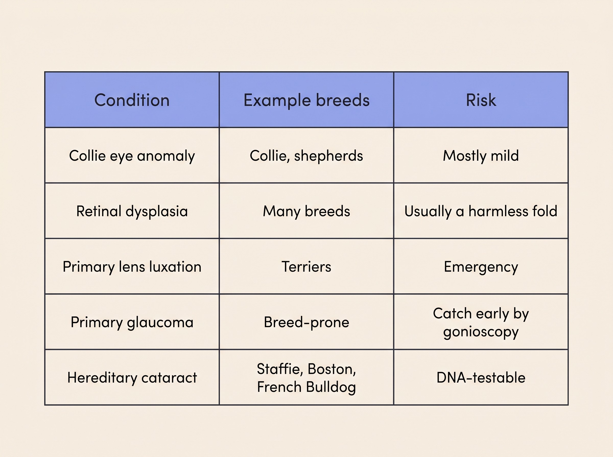
Primary lens luxation: the one emergency on this list

Read this entry carefully, because it breaks the gentle pattern of the others. A sudden, red, painful or cloudy eye in an at-risk terrier is a same-day emergency, not a wait-and-see; if that's happening as you read, use the eye-emergency triage and call a vet now.

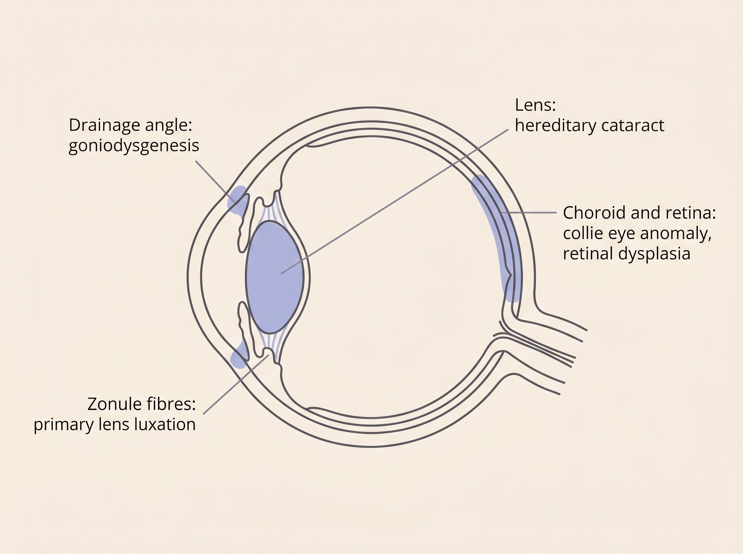
Primary lens luxation (PLL) is an inherited weakening of the zonules, the tiny fibres that hold the lens in place. When they give way the lens slips out of position, and if it drops forward it blocks fluid drainage and triggers a rapid, painful secondary glaucoma that can cause irreversible vision loss if it isn't treated quickly (CAGT, n.d.; Gould et al., 2011). It typically strikes in middle age, around three to eight years, is almost always bilateral (both eyes, often weeks to months apart), and presents as a sudden painful, cloudy, red eye (CAGT, n.d.). The mechanism is glaucoma, but the disease itself, the hours-window and the second-eye risk all belong to glaucoma in dogs; here, just hold the rule that this is the inherited condition where speed saves sight.

It's overwhelmingly a terrier story. The single recessive ADAMTS17 mutation has been found in more than a dozen additional breeds beyond the first three, most of them terriers or breeds with terrier ancestry, including the Jack Russell, Parson Russell, Miniature Bull, Lancashire Heeler, Fox, Tibetan and Yorkshire Terriers and the Chinese Crested (Farias et al., 2010; Gould et al., 2011). One important nuance: although PLL is mostly recessive, carriers with a single copy carry a small but real risk of luxating too, so a carrier of an at-risk breed isn't entirely off the hook (Gould et al., 2011). If you own one of these breeds, knowing the emergency signs is sight-saving knowledge.

Breed-prone primary glaucoma: an inherited narrow drain

Some breeds are born with a malformed drainage angle, a condition called goniodysgenesis or pectinate ligament dysplasia, where abnormal tissue partly blocks the angle through which fluid should leave the eye. This predisposes them to primary, closed-angle glaucoma, a painful, blinding pressure rise (Pearl et al., 2013; Vet Times, n.d.). The valuable part is that the malformation can be found before the pressure ever rises, by gonioscopy, where a special contact lens lets the vet view the drainage angle and grade how much of it is abnormal (grade 1 is 25% or less, grade 2 is 26 to 75%, grade 3 is over 75%, on the scale used from January 2026) (Royal Kennel Club, n.d.; BVA, n.d.). More than 40 breeds have been reported with primary glaucoma worldwide; UK eye-scheme breeds screened by gonioscopy include the Flat-Coated Retriever, Welsh and English Springer Spaniel, American Cocker Spaniel, Basset Hound, Great Dane, Dandie Dinmont Terrier and Siberian Husky (Vet Times, n.d.; BVA, n.d.). One honest update: goniodysgenesis was long thought purely congenital, but it can progress with age in several breeds, the Flat-Coated Retriever among them, which is why current advice is to re-screen periodically, roughly every three years, rather than test once (Pearl et al., 2013; BVA, n.d.). The glaucoma itself, its emergency framing and the plan for the second eye live in glaucoma in dogs and protecting the fellow eye; here, the point is that an inherited narrow angle is a predisposition you can catch early, and an unexplained red, painful eye in a predisposed breed is still a same-day call.

Hereditary cataract: not just diabetes and old age

When most people hear "cataract" they think of the diabetic or the elderly dog, but a number of breeds carry an inherited, or primary, cataract that's neither. It's often juvenile, developing in the first one to three years, and usually bilateral and progressive, though how fast varies a lot between breeds (UFAW/Science for Animal Welfare, n.d.; Mellersh et al., 2006). A well-characterised example is the HSF4-gene cataract of the Staffordshire Bull Terrier, Boston Terrier and French Bulldog, a recessive form often visible by around 12 to 15 months that can progress to blindness, and for which a DNA test exists (Mellersh et al., 2006; UFAW/Science for Animal Welfare, n.d.). Inherited cataract is, in fact, one of the leading causes of cataract in dogs across the breeds, alongside diabetes, age, trauma and inflammation; vets often work on the cautious assumption that a cataract is hereditary unless a clear cause such as diabetes or injury is found (Cornell Riney, n.d.). The practical message is simple: a young dog of a predisposed breed developing a cataract is a candidate for the inherited form, but the eye-health questions and the operate-or-not decision are exactly the same as for any cataract, so I'll hand the full explainer to cataracts explained and the choice to the cataract surgery decision.

A clean reference panel on a cream background pairing each condition with example breeds and a simple risk badge: collie eye anomaly with collie and shepherd breeds marked mostly mild; retinal dysplasia marked usually a harmless fold; primary lens luxation with terrier breeds marked emergency; breed-prone primary glaucoma marked catch it early by gonioscopy; hereditary cataract with Staffordshire Bull Terrier, Boston Terrier and French Bulldog marked DNA-testable
A quick orientation, not a diagnosis. Most of these are mild or catchable early; primary lens luxation is the one that needs a vet the same day.

What about cats?

One honest sentence, because this round-up is genuinely dog-heavy. Named, breed-specific inherited eye disease of this kind is overwhelmingly a dog story; cats have far fewer recognised hereditary eye conditions, and feline cataract and glaucoma are usually secondary to other disease, covered in cataracts in cats and glaucoma in cats.

So what do you actually do with this?

Two threads to leave you holding. First, "inherited" really doesn't mean "doomed": a retinal fold may never matter and can fade, mild collie eye anomaly leaves vision intact for life, a carrier dog is usually perfectly healthy, and even the serious conditions are increasingly detectable early by eye exam, gonioscopy or DNA test, which buys time and choices rather than taking them away.

Second, the concrete thing I'd like you to do. If you have, or are about to buy, one of these breeds, ask whether the parents were eye-tested under the BVA/KC/ISDS scheme, and DNA-tested where a test exists for that breed; and have your own dog screened if your vet advises it. That single question turns "is my breed doomed?" into "here's what's been checked". For the depth, the dedicated pieces are where it lives: progressive retinal atrophy for the big inherited retinal disease, glaucoma in dogs for the painful emergency, the cataract surgery decision for the operate-or-not question, and the breed and DNA-testing piece for what a result actually means. And for any predisposed dog whose sight you simply want to watch over the years, the at-home vision check gives you a repeatable way to catch change early, which with this group of conditions is almost always what helps most. If a slowly progressive condition is on the cards, preparing for blindness is worth a look too, because a long runway is one real advantage these gradual conditions hand you.

References

  1. British Veterinary Association. (n.d.). Eye Scheme (Canine Health Schemes; Known Inherited Ocular Diseases list; gonioscopy grading; breeds screened by gonioscopy). British Veterinary Association.
  2. BVA/KC/ISDS Eye Scheme. (2024). Information for owners. British Veterinary Association, Kennel Club and International Sheep Dog Society.
  3. Canine Genetic Testing (CAGT), University of Cambridge. (n.d.). Primary lens luxation.
  4. Cornell University College of Veterinary Medicine, Riney Canine Health Center. (n.d.). Canine cataracts.
  5. Farias, F. H. G., Johnson, G. S., Taylor, J. F., Giuliano, E., Katz, M. L., Sanders, D. N., Schnabel, R. D., McKay, S. D., Khan, S., Gharahkhani, P., O'Leary, C. A., Pettitt, L., Forman, O. P., Boursnell, M., McLaughlin, B., Ahonen, S., Lohi, H., Hernandez-Merino, E., Gould, D. J., Sargan, D. R., & Mellersh, C. S. (2010). An ADAMTS17 splice donor site mutation in dogs with primary lens luxation. Investigative Ophthalmology & Visual Science, 51(9), 4716-4721.
  6. Gould, D., Pettitt, L., McLaughlin, B., Holmes, N., Forman, O., Thomas, A., Ahonen, S., Lohi, H., O'Leary, C., Sargan, D., & Mellersh, C. (2011). ADAMTS17 mutation associated with primary lens luxation is widespread among breeds. Veterinary Ophthalmology, 14(6), 378-384.
  7. Mellersh, C. S., Pettitt, L., Forman, O. P., Vaudin, M., & Barnett, K. C. (2006). Identification of mutations in HSF4 in dogs of three different breeds with hereditary cataracts. Veterinary Ophthalmology, 9(5), 369-378.
  8. MSD (Merck) Veterinary Manual. (n.d.). The ocular fundus in animals (retinal dysplasia and collie eye anomaly).
  9. Online Mendelian Inheritance in Animals (OMIA). (n.d.). OMIA:000218-9615: Choroidal hypoplasia, NHEJ1-related in Canis lupus familiaris (dog).
  10. Parker, H. G., Kukekova, A. V., Akey, D. T., Goldstein, O., Kirkness, E. F., Baysac, K. C., Mosher, D. S., Aguirre, G. D., Acland, G. M., & Ostrander, E. A. (2007). Breed relationships facilitate fine-mapping studies: A 7.8-kb deletion cosegregates with collie eye anomaly across multiple dog breeds. Genome Research, 17(11), 1562-1571.
  11. Pearl, R., Gould, D., & Spiess, B. (2013). Progression of pectinate ligament dysplasia over time in two populations of Flat-Coated Retrievers. Veterinary Ophthalmology, 18(1), 6-12.
  12. The Royal Kennel Club. (n.d.). Eye screening scheme (including multifocal retinal dysplasia, primary glaucoma and gonioscopy grading).
  13. Srikanth, A., Sukcharoen, P., Boonyawiwat, V., & Yodsheewan, R. (2022). A novel multiplex polymerase chain reaction assay for the genotypic survey of the non-homologous end-joining factor 1 gene associated with collie eye anomaly in Thailand. PMC8924400.
  14. UFAW / Science for Animal Welfare Network. (n.d.). Boston Terrier: hereditary cataract (Genetic Welfare Problems of Companion Animals).
  15. Vet Times. (n.d.). Canine primary glaucoma (clinical review; goniodysgenesis, pectinate ligament dysplasia and primary glaucoma).