What Cataract Surgery Involves, and Recovery Week by Week
Dr. Alastair Greenway
MRCVS
If you've reached this page, you've probably already done the hard part. The agonising over whether to operate, the cost, the "is it the right thing for my dog", that all belongs to the decision, and if you're still weighing it up, the operate-or-not piece is the place for it. What I want to do here is simpler and, I hope, calmer: walk you through exactly what the operation involves and what the weeks afterwards will be like, so that none of it arrives as a surprise.
I'll be honest about one thing up front, because it shapes everything that follows. The surgery itself is over in an afternoon. The recovery is not. The drops, the cone, the quiet weeks and the run of rechecks are real work, and they're the part you can directly influence. The good news is that it's manageable, thousands of owners do it every year, and that careful aftercare is precisely what protects a very good result.
The operation in plain terms
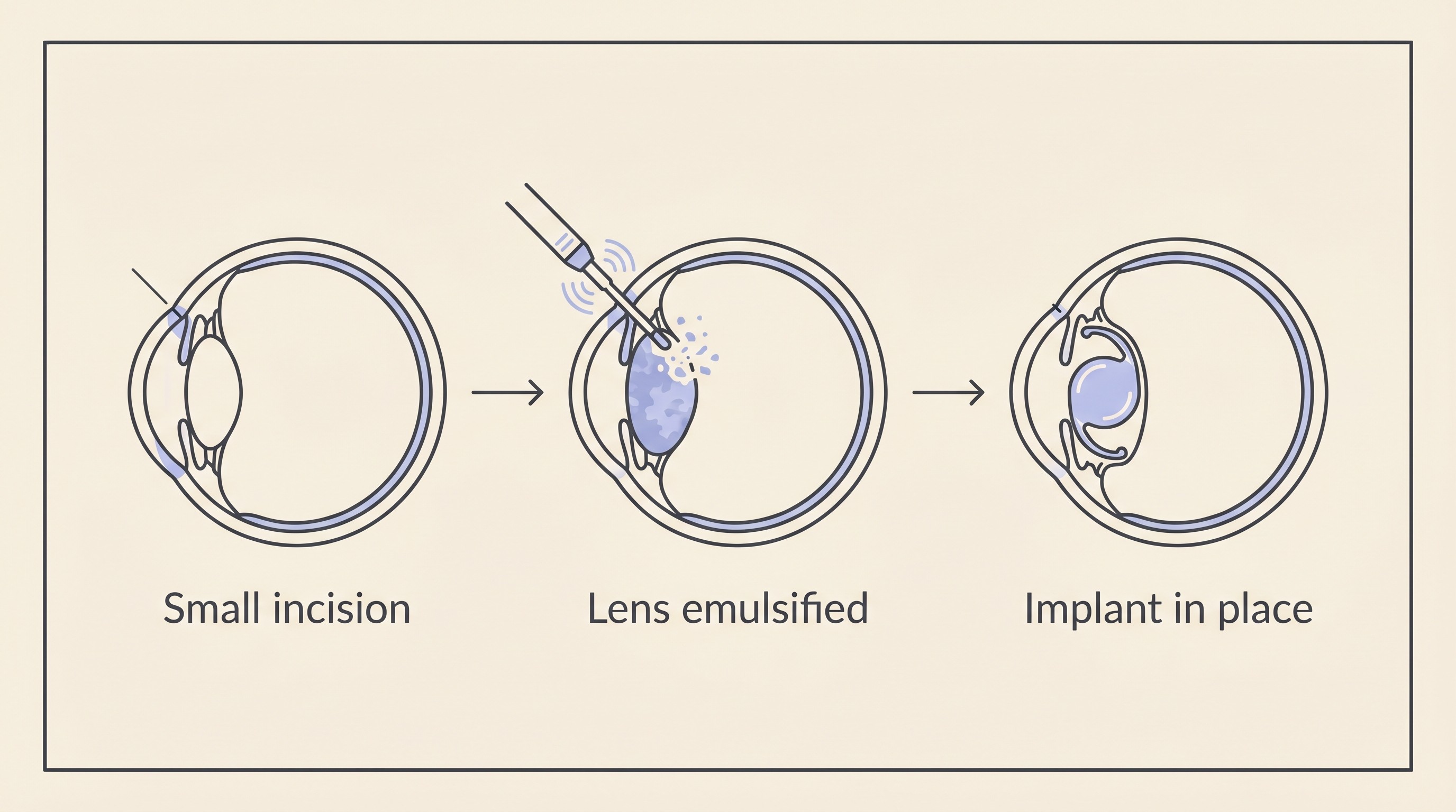
Cataract surgery in pets is phacoemulsification, usually shortened to "phaco", and it's the very same technique used for human cataracts, which owners often find reassuring (Willows, 2024; North Downs Specialist Referrals, 2024). Here's what actually happens. Your pet goes under a general anaesthetic, and the surgeon, working through an operating microscope, makes a tiny incision, only around 3 mm, in the clear cornea at the front of the eye (MSPCA-Angell, 2023). A small window is opened in the capsule that holds the lens, the cloudy lens is broken up by an ultrasonic probe and gently suctioned out, and in most cases an artificial intraocular lens, an IOL, is slipped into the empty capsule to take over the focusing job (Cornell Riney Canine Health Center, 2023; MSPCA-Angell, 2023). The lenses are made specifically for dogs or for cats, because their eyes focus differently from ours (Willows, 2024).
Before any of this, the surgeon has to be sure the retina behind that cloudy lens is healthy, because a mature cataract hides the back of the eye from view. That's done with an electroretinogram (ERG), which checks the retina is still working, and an ocular ultrasound, which rules out a retinal detachment lurking out of sight (MSPCA-Angell, 2023; Willows, 2024; Cornell Riney Canine Health Center, 2023). This candidacy gate lives properly in the decision piece; just know that if your pet has reached the operating list, these checks have already given the green light.
In the UK, most patients stay in overnight and go home the following day once the eye has settled, with a cone already on, a bag of drops and tablets, and the first recheck in the diary (Willows, 2024; North Downs Specialist Referrals, 2024).
How well it works
Let me give you the honest figure, because it's a genuinely good one. Around 90 to 95% of eyes regain useful vision initially after surgery (North Downs Specialist Referrals, 2024; MSPCA-Angell, 2023). That does mean roughly one eye in ten or twenty doesn't get the vision back, and I won't gloss over that, but for most pets this works and works well, with one referral series of 179 eyes finding 82.7% still functionally visual at the end of follow-up (Klein et al., 2011).
Timing nudges that outcome, which is the one part of it you can influence. The ideal time to operate is before a cataract becomes fully mature, because a long-standing, hypermature lens leaks more protein and stirs up more inflammation inside the eye, and that inflammation is what drives the complications that lower a result (Cornell Riney Canine Health Center, 2023; University of Illinois, 2025). By the mature and hypermature stage essentially every eye has some of that lens-induced inflammation, and a hypermature cataract carries a measurably higher risk of glaucoma than an immature one (University of Illinois, 2025; Sigle and Nasisse, 2006). That's why "sooner is better" gets repeated in this space, and why you won't be told to wait and watch a surgical cataract for months. The maturation detail belongs to cataracts explained, and the particular urgency of fast-forming diabetic cataracts to diabetic cataracts.
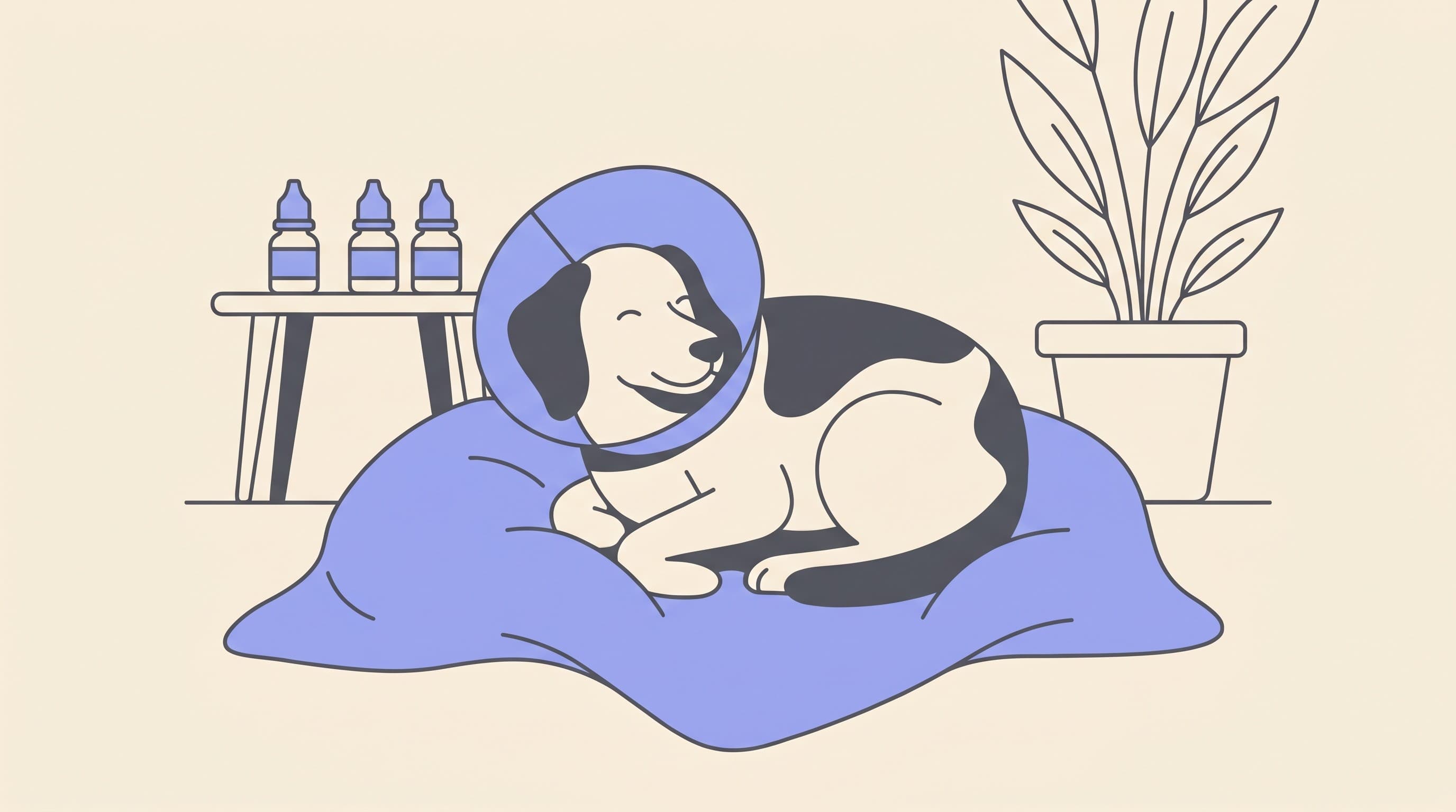
The recovery, week by week
This is the heart of it, and the part most owners want mapped out. Recovery from cataract surgery isn't painful for the pet in the way you might fear, but it is busy, and two things dominate the first couple of months: the eye drops, and protecting the eye. Get those right and you've done the lion's share of the work. One honest caveat before the timeline, though. Every centre runs its own protocol, so the exact drops, the cone duration and the recheck dates vary between surgeons. What follows is the shape of it, anchored to a published UK schedule, not a prescription, so follow your own surgeon's instructions over anything you read here.
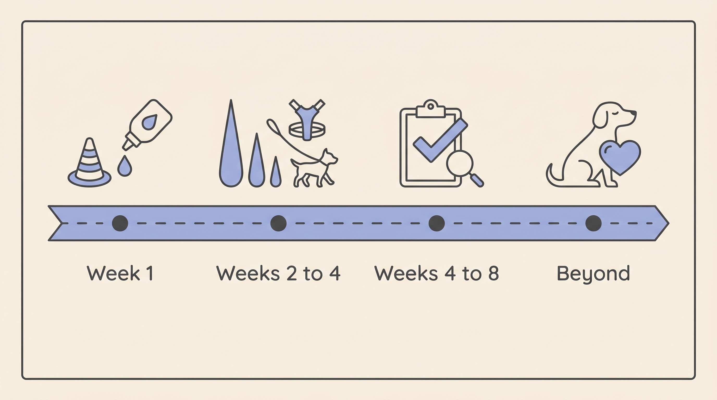
Week 1. The eye will look a little red and may weep, which is expected. Your pet wears a protective collar, a cone or buster collar, around the clock to stop any rubbing or scratching, because a paw or a carpet at the wrong moment can undo delicate work (North Downs Specialist Referrals, 2024; Cornell Riney Canine Health Center, 2023). The drops start at their most intensive, typically four to six times a day, alongside anti-inflammatory tablets (Cornell Riney Canine Health Center, 2023; North Downs Specialist Referrals, 2024; Willows, 2024). Keep life calm: lead walks only, no rough play, no jumping on and off furniture, and crucially walk on a harness rather than anything around the neck, because pulling against a collar raises the pressure inside the eye and can encourage bleeding (North Downs Specialist Referrals, 2024; Willows, 2024; Davies Veterinary Specialists, n.d.). The first recheck usually lands at about a week (Davies Veterinary Specialists, n.d.).
Weeks 2 to 4. Things settle. A published UK schedule gives a sense of the trajectory: drops four times daily for the first four weeks, then three times daily for the next four, then twice daily, and so on, with tablets twice daily for around four weeks (Davies Veterinary Specialists, n.d.). The cone comes off somewhere in this window in many cases, anywhere from about one week to three depending on the centre and how the eye is healing (North Downs Specialist Referrals, 2024; Willows, 2024). Activity stays restricted, harness walks continue, and there's usually a recheck around two weeks and again around four (Davies Veterinary Specialists, n.d.).
Weeks 4 to 8. The drops taper further as the eye proves stable, and your pet gradually returns to normal life on the surgeon's say-so. A recheck around eight weeks is typical, and by this point most eyes are doing the thing you hoped for: seeing. All told, expect at least four or five re-examinations, the bulk in the first two to three months, then settling to every three to six (North Downs Specialist Referrals, 2024; Davies Veterinary Specialists, n.d.).
A word on the drops, because they matter more than anything else you'll do. The post-operative inflammation control is what protects the result, and getting those drops in, on time, every time, is the single biggest factor genuinely within your hands (Cornell Riney Canine Health Center, 2023; Davies Veterinary Specialists, n.d.). A missed dose here or there isn't a catastrophe, but a pattern of missed drops is a real and avoidable risk to the eye. If juggling several drops several times a day sounds daunting, that's exactly what the Eye-Drop and IOP tracker is built for: a per-eye schedule, reminders so a dose doesn't slip, and a place to note how the eye looks, with a printable eye-drop schedule to match. Lining up the bottle caps by colour and having a second person help in the first few days both make it easier than it sounds.
Many eyes also need at least one long-term, sometimes lifelong, topical medication and routine monitoring even after an excellent result (Cornell Riney Canine Health Center, 2023). That's not a sign anything went wrong, just part of looking after an eye that has had surgery, and for most owners it settles into a quick once-a-day habit.
The complications to know, and when to ring
No honest account skips this part. The serious complications to be aware of are raised pressure inside the eye (post-operative ocular hypertension, which can become glaucoma), inflammation inside the eye (uveitis), retinal detachment, bleeding into the eye, corneal ulcers or infection, and posterior capsular opacification, sometimes called "after-cataract" (Sigle and Nasisse, 2006; Klein et al., 2011; Willows, 2024; North Downs Specialist Referrals, 2024).
Honest numbers help more than the bare word "complications", so here are some from referral populations. In a series of 179 eyes, post-operative ocular hypertension occurred in about 22.9%, uveitis in 16.2%, bleeding into the eye in 12.3%, retinal detachment in 8.4% and glaucoma in 6.7% (Klein et al., 2011). A weighted summary across studies puts glaucoma at around 11%, uveitis around 9% and retinal detachment around 4%, with posterior capsular opacification the common one at around 60% (MSPCA-Angell, 2023). That last figure looks alarming until you understand it: "after-cataract" is a clouding of the thin membrane behind the new lens rather than a return of the cataract, usually mild, and it rarely costs vision that matters (MSPCA-Angell, 2023).
The two that genuinely matter, because they can blind the eye, are glaucoma and retinal detachment (MSPCA-Angell, 2023; Sigle and Nasisse, 2006). The reassuring counterweight is that they aren't common and don't tend to strike early: in a 290-eye series, outright failure, an eye that ended up blind or needing removal, stayed below 10% until after three years (Sigle and Nasisse, 2006). So the headline isn't "this is risky", it's "this works well, and here's the small print, told straight".
What does all this mean for you at home? Mostly, knowing the signs that say "be seen today". If the operated eye suddenly becomes red, cloudy or very painful, or your pet seems to lose vision in it, that's not a wait-and-see: those are the signs of a pressure spike, a flare of inflammation or a detachment, and they need an urgent call to your surgeon or an emergency vet the same day (Willows, 2024; North Downs Specialist Referrals, 2024). Squinting, pawing at the eye, holding it shut or going quiet and off food all count as pain until proven otherwise. If you're ever unsure how urgent something is, the eye-emergency triage will help you sort a today problem from a routine one. And in the rare event a complication does cost the eye its vision, pets adapt remarkably well, so the living-with cluster is there rather than any cause to catastrophise.
A note for cat owners, and the cost
This piece is written for both species, but cataract surgery is overwhelmingly a dog operation. In cats, cataracts are uncommon and usually secondary to long-standing inflammation inside the eye, so the underlying disease is treated first and surgery is reserved for selected cases. The operation and recovery follow the same principles, but the feline picture has its own shape, and cataracts in cats is the place for it.
On cost, one honest line, because the full discussion sits with the decision piece: UK cataract surgery runs roughly £3,500 to £5,000 per eye as a practical estimate, varying by centre and by whether one eye or both are done (Eye-Vet, 2024).
So here's where I'd leave you. The operation is quick and refined, the success rate is genuinely good, and the recovery, busy as it is, follows a clear and predictable shape that thousands of owners navigate every year. Your job in it is concrete and doable: get the drops in on time, keep the eye protected, keep life calm, and turn up to the rechecks. If you haven't yet had the pre-operative consult, take the questions worth asking with you, or print the consult question list, so you leave knowing your own pet's odds and schedule rather than the averages on this page. And if you set up one practical thing before the day itself, make it the drop tracker, loaded with your surgeon's schedule, so that from the moment you walk back through your own front door the hardest part of the aftercare is already organised.
References
- Cornell University College of Veterinary Medicine, Riney Canine Health Center. (2023). Canine cataracts.
- Davies Veterinary Specialists. (n.d.). Owner information sheet for cataract surgery.
- Eye-Vet (Runcorn, UK). (2024). Price list: cataract surgery.
- Klein, H. E., Krohne, S. G., Moore, G. E., & Stiles, J. (2011). Postoperative complications and visual outcomes of phacoemulsification in 103 dogs (179 eyes): 2006-2008. Veterinary Ophthalmology, 14(2), 114-120.
- MSPCA-Angell (Angell Animal Medical Center), Martin, K. (2023). Lifting the veil: canine cataracts and cataract surgery.
- North Downs Specialist Referrals (NDSR). (2024). Veterinary cataract surgery.
- Sigle, K. J., & Nasisse, M. P. (2006). Long-term complications after phacoemulsification for cataract removal in dogs: 172 cases (1995-2002). Journal of the American Veterinary Medical Association, 228(1), 74-79.
- University of Illinois College of Veterinary Medicine. (2025). Cataracts in the small animal patient.
- Willows Veterinary Centre and Referral Service. (2024). Cataract surgery.
Keep track of how your pet is doing
The owners who cope best are the ones who notice changes early. A simple health log shows you what is working, and what is not, before the next vet visit.
Start tracking, freeYou're not doing this alone
Compare treatment journeys and talk to owners managing vision & eye health. Free to join.
Join PetsLikeMine