If You Don't Operate: Living With a Pet With Cataracts
Claire Greenway
BVM&S MRCVS
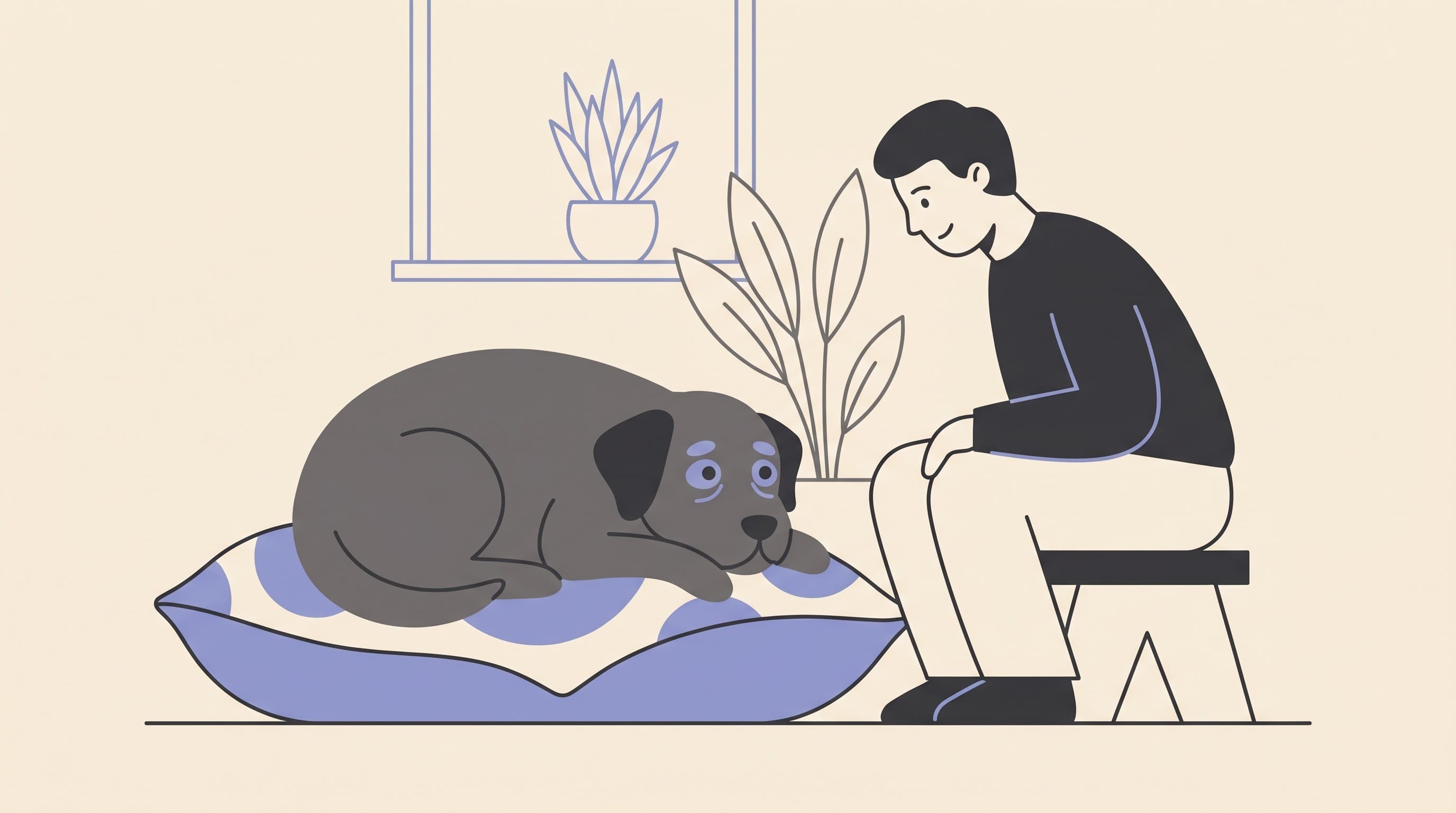
Plenty of owners arrive at this decision quietly, and often with a flicker of guilt. The surgery is specialist, it isn't cheap, your pet may be older or not the ideal candidate, and somewhere in the back of your mind sits the worry that by not operating you're "letting them go blind". That worry deserves an honest answer rather than a pat one.
Choosing not to operate on a cataract is a legitimate, kind choice, not neglect. The cloudiness itself does not hurt, and many pets live perfectly good lives with a cataract, especially when it changes slowly and they have a familiar, settled home to navigate (Cornell Riney, n.d.). The decision itself belongs to its own conversation, and if you're still weighing it up the surgery decision is the place to do that. This article assumes you've chosen not to operate and now want to do right by a pet living with a cataract. There is a real job here, and it's a doable one.
First, the honest reassurance
A cataract is the lens going from clear to cloudy, blocking light and dimming or removing sight in that eye. On its own, that clouding is not painful and not an emergency. So the starting point is genuine reassurance: this is not a crisis, and your pet is not suffering simply because an eye has gone cloudy.
The evidence backs that up more directly than you might expect. In a study comparing cataractous eyes that had surgery with eyes that did not, the owners who simply elected against surgery for an otherwise reasonable eye saw a long-term complication rate of just 3.3%, that's one eye in thirty, and whether or not a cataractous eye had surgery did not change the long-term complication rate overall (Krishnan et al., 2020).
I do have to be straight about one nuance, because it's the honest part. That low 3.3% figure is for eyes that were reasonable surgical candidates whose owners chose not to go ahead. Eyes that were poor candidates in the first place fared less well, with a complication rate of about a third (33%) (Krishnan et al., 2020). So "we decided not to operate on a healthy-but-cloudy eye" is a very different starting hand from "this eye already had other problems". Most pets in the first group do well, and which group your pet is in is a fair thing to ask your vet, because it shapes how closely you'll want to watch.
The real job: watching for the painful complications
Here is the part that matters most, and the reason this article exists. A cataract is more than a cosmetic cloud, because the lens can drive painful inflammation inside the eye, and that's what the no-surgery owner is signing up to watch for.
As a cataract advances, it starts to leak lens proteins into the eye. The immune system has never normally "seen" these proteins, treats them as foreign and attacks, producing what we call lens-induced uveitis (ACVO, n.d.; Maggio, 2015). This isn't a rare curiosity. Lens-induced uveitis is the most common complication of an untreated cataract, found in as many as 71% of eyes examined before cataract surgery, and it's more active in younger animals and in rapidly progressing cataracts, including diabetic ones (Maggio, 2015; ACVO, n.d.).
Left unchecked, that inflammation has a sting in its tail. Lens-induced uveitis is the leading cause of secondary glaucoma in dogs, responsible for around 81% of those cases, and overall almost 20% of dogs with cataracts are reported to develop glaucoma (Maggio, 2015). Glaucoma, a damaging rise in the pressure inside the eye, is painful and sight-threatening, and that is the complication we're really guarding against. The disease itself, and why it's an emergency, is covered in glaucoma in dogs; the wider picture of inflammation inside the eye lives in uveitis. For now, hold the chain of events: cataract, then lens-induced uveitis, then, if it runs unchecked, secondary glaucoma.
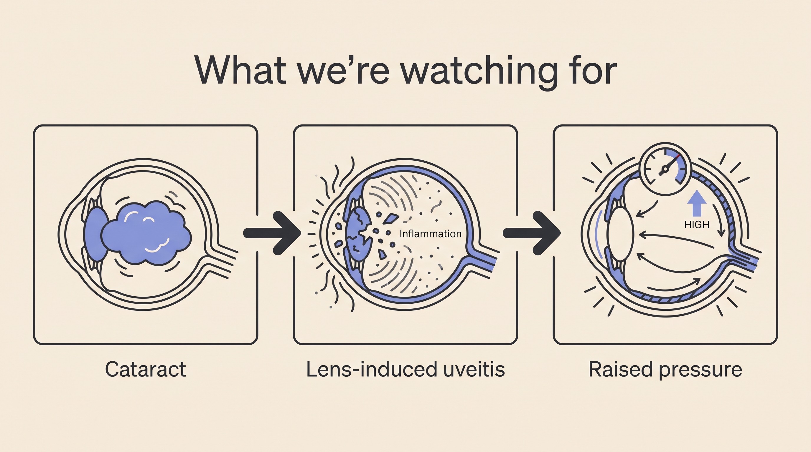
The good news sits right alongside that, and it's why monitoring is worth doing rather than worrying about. Lens-induced uveitis can usually be controlled with topical eye drops, and a corticosteroid or anti-inflammatory drop is the treatment of choice, started promptly once the inflammation is diagnosed (ACVO, n.d.; Allbaugh, 2019). Caught early and treated, the complication is manageable. The case we most want to avoid is a sudden rupture of the lens capsule, most often in a rapidly swelling cataract, which floods the eye with lens material and can drive an inflammatory response that medicine can't control, frequently ending in secondary glaucoma and eye removal (ACVO, n.d.). That's the scenario early vigilance is designed to head off.
The monitoring plan, in plain terms
So what does "watching" actually look like? It's concrete, and it's not onerous. For a pet whose cataract is being managed rather than removed, the standard advice is twofold: talk to your vet about topical anti-inflammatory drops to try to prevent cataract-associated eye disease, and have the eye's pressure checked for glaucoma every four to six months (Cornell Riney, n.d.).
That pressure check is the quiet hero of the plan. Secondary glaucoma can build silently before it ever announces itself as a red, painful crisis, so a routine reading catches it while it's still treatable. In an eye that's actively inflamed, the intraocular pressure should sit below roughly 10 to 15 mmHg, and a higher reading should raise suspicion of secondary glaucoma (Allbaugh, 2019). It's a quick, well-tolerated test, and the single most useful thing a check-up gives you between now and any red flag.
If your vet does start anti-inflammatory drops, the Eye-Drop & IOP tracker keeps the schedule and a home pressure log in one place, and the eye-drop schedule is a printable companion. Between visits, the At-Home Vision Check lets you trend your pet's functional sight, so a change is a fact you've recorded rather than a feeling you're second-guessing.
This cadence isn't over-caution. When lens complications are caught late, eyes and sight are genuinely lost. In one referral series of ninety eyes presenting at a late stage, secondary glaucoma accompanied the great majority of the lens-luxation cases (around 78%) and over a third of the lens-induced-uveitis ones (about 38%), vision was lost in most of the worst-affected eyes, and the authors' conclusion was blunt: early diagnosis and timely treatment are crucial to keeping vision and the eye (Ali & Mostafa, 2023). I don't quote that to frighten you, because those were eyes that presented late, often with problems beyond a simple cataract. I quote it because it's exactly the outcome a four-to-six-month check and a quick call about the red flags are there to prevent.
The red flags: when a routine check becomes a today visit
This is the one part of the article to commit to memory. Most of the time your pet's cloudy eye will tick along quietly. But if a cataract eye suddenly turns:
- red, or noticeably cloudier than usual;
- painful, which in pets means squinting, holding the eye shut, rubbing or pawing at it, going quiet or off their food rather than crying out;
- or suddenly more blind, bumping about when they'd been managing,
then it needs to be seen that day. That picture is lens-induced uveitis flaring or secondary glaucoma setting in, and glaucoma is a sight-threatening emergency measured in hours, not days (Cornell Riney, n.d.; Maggio, 2015). If you're unsure how urgent something is, the eye-emergency triage will sort it for you, and the eye-emergency red-flags sheet is worth sticking on the fridge. Everything else in this plan, the drops, the pressure checks, the steady home, exists to keep this from happening, but if it does, acting the same day is what protects the eye.
Not every cataract changes at the same pace
Part of monitoring is watching the cataract itself, because they don't all progress alike. Across cataracts of different causes, complications are diagnosed in roughly 43.5% overall and become more likely the further the cataract has developed, while the rate of progression varies by cause: age-related cataracts progress in about 30% of cases, hereditary, congenital and diabetic ones far more often, and diabetic, traumatic, secondary and hereditary types carry more complications than age-related ones (Fischer & Meyer-Lindenberg, 2018).
In practice, a slowly-changing age-related cataract in an older dog and a fast-swelling diabetic cataract are two different monitoring jobs. Diabetic cataracts deserve a special flag: most diabetic dogs develop them, around 80% within the first year or so whatever the glucose control, and they can cloud and swell fast (Beam et al., 1999; Cornell Riney, n.d.). A fast, swelling cataract is more prone to the very complications above, and it is exactly the kind most likely to rupture its capsule, so a diabetic dog's eyes warrant closer watching and earlier action, not a comfortable "just monitor". The speed, the referral timing and the guilt-relief diabetic owners need are covered in full in diabetic cataracts, with the glucose-control side in the Diabetes space. If your pet is diabetic, read that next.
A quick word on cats, because they're different. Cataracts are uncommon in cats, and when they do appear they're usually secondary to long-standing inflammation inside the eye rather than primary or diabetic. So a cat with a cataract really needs the underlying cause hunted down and treated, and that search often is the management job. The same red flags and pressure-monitoring logic apply, but the feline picture has its own shape, set out in cataracts in cats.
Helping a pet who can't see as well
If your pet's sight is fading in the affected eye, the home side of this is more reassuring than owners fear. Pets adapt remarkably well to losing vision, especially when it happens gradually. As long as any underlying disease is managed, most dogs adjust well to blindness over about six to eight weeks, and owners who worry the early withdrawn or clingy behaviour is permanent are usually pleasantly surprised that it isn't (RSPCA, n.d.; Weir & Barnette, n.d.). They get about by a memorised map of the house plus scent and sound, which is why a familiar, settled home matters so much.
The single biggest thing you can do is keep that mental map intact. Don't move the furniture, keep food and water bowls and beds where they've always been, and talk before you touch so a low-vision pet isn't startled by a hand they didn't see coming (RSPCA, n.d.; VIN/Veterinary Partner, n.d.). That's the headline. The room-by-room detail, the texture and scent cues, the stairs and gates, all of it is handled properly in home-proofing for a blind pet and the rest of the living-with cluster, and the blind-pet home checklist is a good place to start if you'd like a list in your hand.
The door stays open
Here's the thing I most want you to take away: deciding not to operate now is not a door that locks behind you. If your pet's vision loss accelerates and the eye is otherwise healthy, surgery can be revisited. The honest caveat is that, in general, the sooner a cataract is removed the better, because over time the lens hardens and the risk of those painful secondary complications climbs (Cornell Riney, n.d.; Maggio, 2015). So if circumstances change, don't assume the moment has passed. Have the conversation, and the surgery decision will help you reframe it.
For now, your no-surgery plan is simple and within reach: control any inflammation with the drops your vet advises, have the eye's pressure checked every four to six months, know the red flags, keep the home steady, and keep the surgical option in view in case the picture shifts. That's not second-best medicine. It's a real, active plan that keeps the eye comfortable and your pet themselves, and it's one you can do well.
References
- Allbaugh, R. A. (2019). Managing uveitis in dogs and cats. Today's Veterinary Practice.
- Ali, K. M., & Mostafa, A. A. (2023). Lens-related ocular emergencies (LROE) in dogs: treatment and visual outcome after late presentation of 90 eyes. Irish Veterinary Journal, 76, 12.
- American College of Veterinary Ophthalmologists (Sigle, K.). (n.d.). Uveitis in dogs and cats. ACVO Public.
- Beam, S., Correa, M. T., & Davidson, M. G. (1999). A retrospective-cohort study on the development of cataracts in dogs with diabetes mellitus: 200 cases. Veterinary Ophthalmology, 2(3), 169-172.
- Cornell University College of Veterinary Medicine, Richard P. Riney Canine Health Center. (n.d.). Canine cataracts.
- Fischer, M. C., & Meyer-Lindenberg, A. (2018). Progression and complications of canine cataracts for different stages of development and aetiologies. Journal of Small Animal Practice, 59(10), 616-624.
- Krishnan, H., Hetzel, S., McLellan, G. J., & Bentley, E. (2020). Comparison of outcomes in cataractous eyes of dogs undergoing phacoemulsification versus eyes not undergoing surgery. Veterinary Ophthalmology, 23(2), 286-291.
- Maggio, F. (2015). Canine cataracts: classification, aetiology and complications. Veterinary Times.
- RSPCA. (n.d.). 9 tips for living with blind dogs.
- VIN / Veterinary Partner. (n.d.). Living with blind dogs and cats.
- Weir, M., & Barnette, C. (n.d.). Sudden acquired retinal degeneration syndrome (SARDS). VCA Animal Hospitals.
Keep track of how your pet is doing
The owners who cope best are the ones who notice changes early. A simple health log shows you what is working, and what is not, before the next vet visit.
Start tracking, freeYou're not doing this alone
Compare treatment journeys and talk to owners managing vision & eye health. Free to join.
Join PetsLikeMine