


Hot spots: the angry patch that appears overnight
Alastair Greenway
BVM&S MRCVS
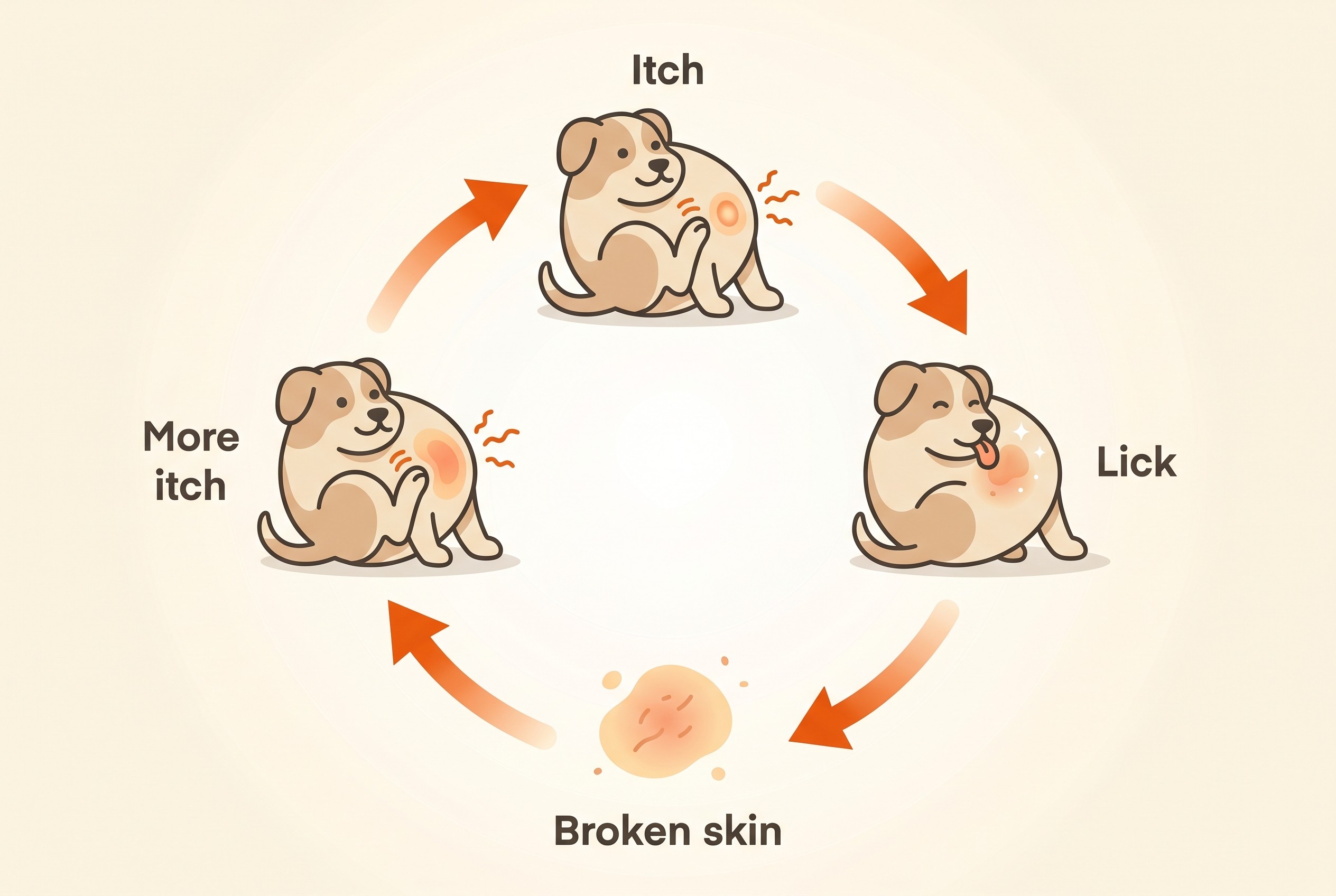
A normal dog goes to bed, and by morning there's a raw, weeping, hairless patch the size of a coin, hot to the touch and clearly sore. It looks alarming, and it genuinely was not there yesterday. The single most useful thing to understand is this: your pet made it, very fast, by licking, chewing or scratching one spot raw because that spot was itchy or sore (Holm et al., 2004; Hardy, 2017). The angry patch is the visible end of a hidden problem, not the problem itself, and that reframes both what you do about it tonight and how you stop the next one.
What a hot spot actually is
The proper name is acute moist dermatitis, also called pyotraumatic dermatitis. Break that second word apart and it tells the story: "pyo-traumatic" means pus-and-trauma (Holm et al., 2004). The skin goes from normal to angry within hours because the damage is mechanical. A dog can chew a spot raw in one afternoon, which is why it feels as though it appeared overnight (Hardy, 2017).
This is the part the content mills get wrong. In the common form, the bacteria are not the root cause. Pyotraumatic dermatitis is classed as a surface pyoderma, where bacteria colonise the broken skin rather than truly infecting it, and there is genuine professional debate about how big a role they play at all (Loeffler et al., 2025; Miller, Griffin and Campbell, 2013). When a swab is taken, what grows is the pet's own normal skin staph: Staphylococcus (then S. intermedius, now reclassified as S. pseudintermedius) grew in 25 of 27 cultured cases in the landmark series (Holm et al., 2004). That is resident flora multiplying on a raw surface, not an infection caught from elsewhere, and not a sign you did anything wrong. The scratching comes first, the bacteria move in afterwards. The general biology of pyoderma, and why it is a secondary problem, lives in skin and ear infections.
Why so fast, and why the site is a clue
Some pets are simply more prone. Hot spots turn up most in dogs with thick, dense, heavy coats, and the best prospective series found Rottweilers, German Shepherds and Golden Retrievers the commonest breeds (Holm et al., 2004). A warm, humid, poorly ventilated coat raises the risk, which is why they often follow a swim, a rained-on walk or a bath that never fully dried. One caveat, because it is repeated everywhere as fact: the "hot spots are a summer thing" line is a pattern many vets see, but that same study, in a flea-free population, found no significant seasonal incidence (Holm et al., 2004). Treat humidity as a risk factor, not an iron law.
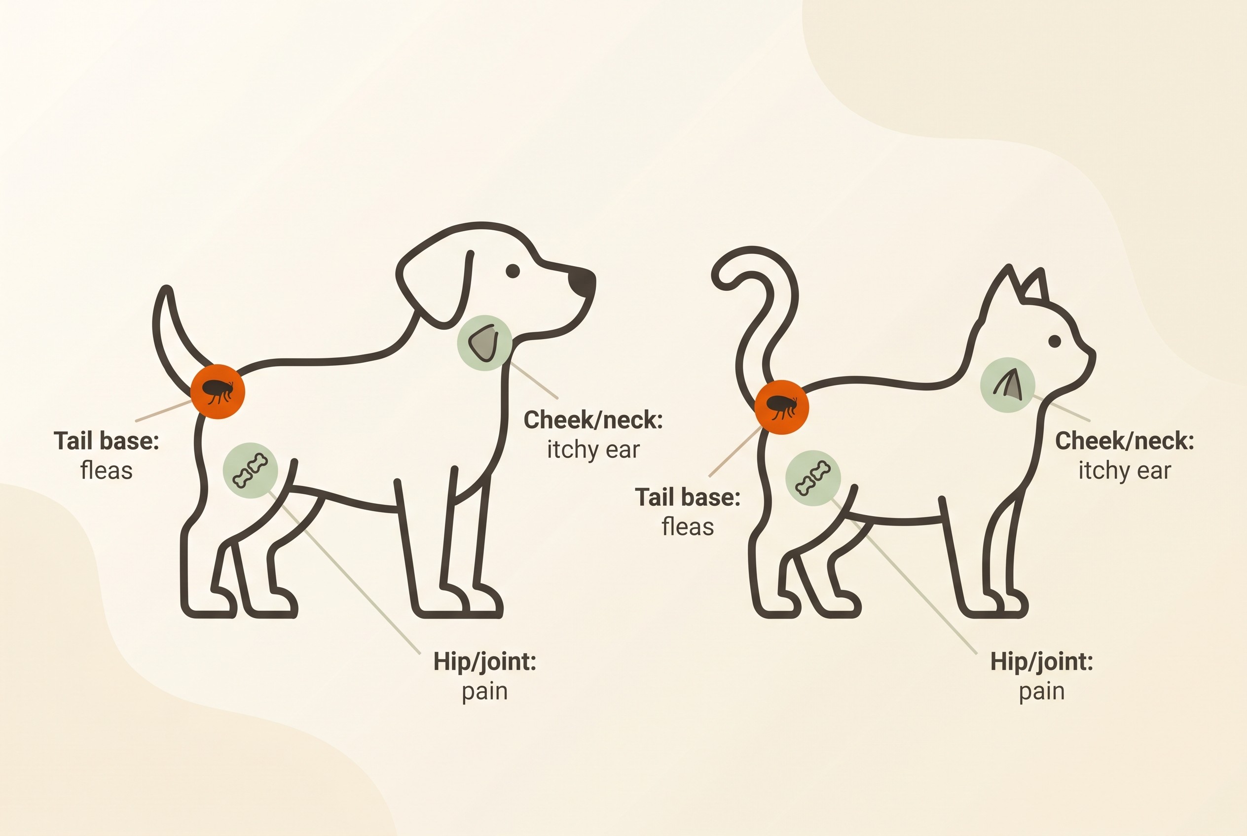
The more useful clue is where the patch lands, because the site often points straight at the trigger. A hot spot at the tail base or rump points hard at fleas, the classic flea-allergy distribution (Holm et al., 2004), and that story is owned by flea allergy dermatitis. One on the cheek, side of the face or neck often sits over an itchy ear the pet has been clawing at, the cue to read recurrent ear infections. A patch over a hip or other joint can mean pain the pet keeps licking. The map gives you a head start on the real question, which is not "how do I treat the patch" but "what set my pet itching".
Surface or deeper: the difference that changes treatment
Here is the clinical heart of the matter. Not all hot spots are the same depth. The common one is a surface lesion (pyotraumatic dermatitis) that clears with topical care alone. But a real minority are deeper: pyotraumatic folliculitis and furunculosis, where bacteria are properly infecting the hair follicles, and those behave like a deep pyoderma that needs systemic antibiotics (Hardy, 2017; Loeffler et al., 2025). This is not a rare technicality: in that same series, acute folliculitis was present in 20 of 44 lesions on biopsy (Holm et al., 2004). So "it is just a surface graze" is not always true.
The tell, in plain language, is "satellite lesions". The vet clips the hair well beyond the obvious patch and examines the skin around it. If it is clean, it is a surface hot spot. If there are little red bumps, pustules or crusts dotted in the seemingly-normal skin nearby, sometimes with thickening or swelling, that signals follicular involvement and a different treatment, and the international guideline is explicit that this peripheral skin should be checked for those signs of deep pyoderma (Loeffler et al., 2025). That is why your vet clips so much wider than the wound. Even experienced vets cannot always tell surface from deep on appearance alone, which is why a lesion that will not settle on topical care earns a recheck rather than a guess, and may have been the deeper kind all along (Hardy, 2017; Holm et al., 2004).
First aid at home, and when it has to be the vet
For a small, early patch the pet will leave alone, there are a few things you can safely do while you arrange a visit. Gently clip or trim the hair over and around it so air can reach the skin and the discharge does not mat, because the wound heals faster dry and open. Clean it with a dilute antiseptic such as chlorhexidine. And above everything else, stop the licking and chewing: every lick re-traumatises the wound and feeds the cycle straight back into itself, so an Elizabethan collar (the cone) or a body suit is the most reliable way to break the loop (Hardy, 2017). No trial proves a cone speeds healing, but you cannot heal a wound the pet keeps reopening. Chlorhexidine is the antiseptic to name because modern guidelines prioritise it over topical antibiotics and there is no meaningful resistance to it (Loeffler et al., 2025; Santoro, 2023).
A few safety points, because owners reach for the bathroom cabinet. Do not use human creams without veterinary advice: many are harmful to pets or simply get licked off. Some "natural" favourites are worse: concentrated tea tree oil, at the strengths people use, is genuinely toxic to dogs and cats and can cause tremors, weakness and collapse (Khan, McLean and Slater, 2014). Do not pick at crusts or put stinging products on raw skin.
Skip home care and book the vet now if the patch is large, spreading or there is more than one; if it is very painful (the pet snaps, cries, or will not let you near it); if it is deep, thickened, or weeping pus rather than clear fluid; if there are satellite spots around it; if your pet is flat, off food or feverish; or if it sits on the face, near an eye, or anywhere you cannot stop the licking. A hot, rapidly spreading or whole-body picture is a different and more urgent problem (an abscess, cellulitis or a severe reaction): that triage belongs to pet skin emergencies, and if it is what you are seeing, go now. If you are unsure whether to home-treat a small patch or book, the itchy pet check can help you decide.
What the vet does, and why finding the trigger is half the visit
The vet is doing two jobs at once: settling the wound and finding what caused it. For the wound, they clip widely (therapeutic, not cosmetic: it lets the skin dry and reveals any satellite lesions), clean with antiseptic, and use a topical anti-inflammatory, usually a glucocorticoid, to break the itch-and-inflammation cycle (Loeffler et al., 2025; Hardy, 2017). Clipping a raw patch genuinely hurts, so many pets need pain relief or light sedation, which is normal, not a sign things are dire (Hardy, 2017).
It is worth knowing how well any of this works. In a randomised, double-blind trial, a topical antimicrobial-plus-corticosteroid combination cleared lesions fastest and a steroid alone worked least well, but the striking detail is that every group, including the untreated controls, had recovered completely within seven days (Schroeder et al., 1996). So surface hot spots tend to settle quickly with basic care, and the anti-inflammatory, not an antibiotic, does much of the heavy lifting. Hence a headline we can state with authority: for a true surface hot spot, oral antibiotics are usually not needed, and topical care is the treatment of choice, because the surface is so directly accessible to it (Loeffler et al., 2025; Santoro, 2023). Tablets are reserved for the deeper, satellite-lesion kind (Holm et al., 2004). So if a vet reaches straight for "have some antibiotics", it is fair to ask whether this is the surface or deep kind; the full topical-first stewardship argument is in treating skin infections.
The second job, finding the trigger, is what stops you being back here in two months. Because the hot spot is driven by an itch or pain, the vet looks for the cause behind it: fleas, an underlying allergy, an ear infection, a sore joint, a matted damp coat (Holm et al., 2004; Loeffler et al., 2025). Treating that cause, not just the patch, is the whole game. The five-cause itch differential belongs to why is my pet itchy, and the way several small triggers stack up to tip a pet over its itch threshold explains why the flare arrived this week and not last.
A note on cats
True hot spots are uncommon in cats. When a cat does present with a moist, eroded, well-circumscribed patch that an owner reads as a hot spot, it is more often an eosinophilic plaque, an allergy-driven lesion that looks similar but is a different beast. The underlying causes are the same family as in dogs (flea allergy, atopy, food allergy and anxiety-driven overgrooming), but the feline picture has its own patterns and is owned by feline atopic syndrome, the article to read if your cat is affected.
Stopping the next one
Prevention follows directly from the cause, so think of it as treating the trigger, not the patch. Rock-solid, year-round flea control is the single highest-yield move for tail-base and rump hot spots (flea control that works). Getting an underlying allergy managed so the itch stays below the threshold is the lasting fix for allergy-driven patches (atopic dermatitis treatment); the allergy drugs you may have heard of, oclacitinib (Apoquel), lokivetmab (Cytopoint, licensed in dogs) and ciclosporin (Atopica), control that underlying itch, not the hot spot, so they belong to that plan, not this one. Treating recurrent ear disease before the dog claws its cheek raw, and drying dense coats thoroughly and clipping out mats so the skin can breathe, round it out (Holm et al., 2004).
A hot spot that keeps coming back is not bad luck, it is a message: a pet that gets two or three a year is telling you its itch, or its fleas, or its ears, is not yet under control. This is where the Skin & Itch Tracker earns its place. Logging where each patch lands, how often, and what was going on at the time (after a swim, mid-pollen-season, a flea spotted), with a photo and an itch score each time, turns "another hot spot" into a pattern you and your vet can act on, and that is how you get to fewer of them.
References
- Holm BR, Rest JR, Seewald W. "A prospective study of the clinical findings, treatment and histopathology of 44 cases of pyotraumatic dermatitis." Veterinary Dermatology. 2004 Dec;15(6):369-376. — WEB-VERIFIED (full abstract read verbatim from PubMed). Confidence: HIGH. The single best primary source for this article. Supports: hot spot = pyotraumatic dermatitis; self-trauma pathogenesis; males > females and dogs <=4 years more affected; commonest sites cheek, neck and lateral thigh with significant breed-by-site correlation; commonest breeds Rottweiler, German shepherd, golden retriever; a possible underlying cause found/suspected in 31/44; NO significant seasonal incidence (the honesty point on the "summer" myth); four histological patterns by eosinophils and/or folliculitis; acute folliculitis present in 20/44 (the surface-vs-deep point); Staphylococcus intermedius (now S. pseudintermedius) grown in 25/27 cultures. URL:.
- Loeffler A, Cain CL, Ferrer L, et al. "Antimicrobial use guidelines for canine pyoderma by the International Society for Companion Animal Infectious Diseases (ISCAID)." Veterinary Dermatology. 2025;36(3):234-282. — WEB-VERIFIED (key passages read via PMC open-access full text). Confidence: HIGH. The current authoritative consensus. Supports: classification of pyoderma into surface/superficial/deep, with pyotraumatic dermatitis as a SURFACE pyoderma; topical antimicrobial therapy is the treatment of choice for surface pyoderma; "systemic antimicrobial therapy should be avoided in cases of surface pyoderma given the location and direct accessibility of microbial pathogens"; chlorhexidine antiseptics prioritised over topical antibiotics; no clinically meaningful chlorhexidine resistance described; the instruction to clip and examine surrounding skin for signs of deep pyoderma; peripheral "papules, pustules, haemorrhagic crusts, swelling and furuncles... indicate... deep pyoderma requiring a different treatment approach" (the satellite-lesion / when-systemic-antibiotics-are-justified point). URL (open access): · Journal DOI:.
- Schroeder H, Swan GE, Berry WL, Pearson J. "Efficacy of a topical antimicrobial-anti-inflammatory combination in the treatment of pyotraumatic dermatitis in dogs." Veterinary Dermatology. 1996;7(3):163-170. — WEB-VERIFIED (citation and abstract confirmed via PubMed record, which indexes this 1996 paper). Confidence: HIGH. A genuine randomised controlled treatment trial in pyotraumatic dermatitis. Supports: clip-and-clean before topical treatment; an antimicrobial-plus-corticosteroid combination gave fastest recovery, antimicrobial alone next, corticosteroid alone poorest; and the honest, quotable detail that ALL groups, including untreated controls, recovered completely within 7 days (ie surface hot spots often settle quickly with basic care; treatment buys comfort and speed). NOTE for drafter: PubMed lists this under with a confusing modern index date, but the paper itself is Vet Dermatol 1996;7(3):163-170. Cite it as 1996. URL:.
- Santoro D. "Topical therapy for canine pyoderma: what is new?" Journal of the American Veterinary Medical Association. 2023;261(S1):S140-S148. — WEB-VERIFIED (author, citation and stewardship message confirmed via PubMed. Confidence: HIGH. Supports: for superficial pyoderma, topical antibacterials alone can be as effective as systemic therapy if frequency, contact time, duration and active ingredient are right and the underlying cause is treated; chlorhexidine (with benzoyl peroxide and ethyl lactate) is a long-used low-resistance topical; systemic antibiotics should be limited to severe cases to reduce selection for multidrug-resistant bacteria. Use for the "topical-first, antibiotics are not the reflex" stewardship line (then hand the deep dive to 4.4). URL: · Full text:.
- Borio S, Colombo S, La Rosa G, De Lucia M, Damborg P, Guardabassi L. "Effectiveness of a combined (4% chlorhexidine digluconate shampoo and solution) protocol in MRS and non-MRS canine superficial pyoderma: a randomized, blinded, antibiotic-controlled study." Veterinary Dermatology. 2015 Oct;26(5):339-344. — WEB-VERIFIED (citation and conclusion confirmed via PubMed). Confidence: HIGH. Supports the broader topical-chlorhexidine evidence base (resolution comparable to systemic amoxicillin-clavulanate, including MRSP cases). Use only as a one-line backstop for "chlorhexidine works and reduces the need for tablets"; the full topical-vs-systemic argument is owned by `treating-skin-infections` (4.4). URL: · Journal:.
- Hardy J. "Dermatology: hot spots in dogs - treatment and advice for owners." Veterinary Times (clinical CPD). 2019. — WEB-VERIFIED (full clinical article read). Confidence: MEDIUM-HIGH (peer-reviewed-grade veterinary CPD trade journal rather than an indexed primary study; content fully consistent with Holm 2004 and ISCAID 2025). Supports: the explicit clinical distinction between surface pyotraumatic dermatitis (colonised, not a true infection) and pyotraumatic folliculitis/furunculosis (true follicular pyoderma); satellite papules/pustules as the marker of follicular involvement, often only visible once clipped; differentiation can be difficult on clinical signs alone; clip and clean with chlorhexidine; clipping/cleaning is painful so sedation may be needed; topical glucocorticoid preferred over systemic for a focal lesion; systemic antibiotics usually NOT needed unless skin thickening and satellite lesions are present; success depends on identifying the underlying trigger. URL:.
- MSD Veterinary Manual (Merck Veterinary Manual). "Pyoderma in Dogs and Cats" / "Feline Atopic Skin Syndrome." Integumentary System. Accessed June 2026. — PARTIALLY WEB-VERIFIED (the pyotraumatic-dermatitis sub-page and the feline atopic skin syndrome page were 403-blocked to direct fetch; content corroborated via search snippets and cross-checked against the primary literature above). Confidence: MEDIUM for direct quotation, HIGH for the facts it backs because they are independently supported by Holm 2004 and ISCAID 2025. Supports (as a textbook-grade backstop, not a sole source): S. pseudintermedius as the normal resident staph; self-trauma pathogenesis; rapid onset; breed/coat predisposition; site-as-clue; and the four feline cutaneous reaction patterns (self-induced alopecia, miliary dermatitis, head and neck pruritus, eosinophilic granuloma complex) plus secondary pyoderma in areas of feline self-trauma. URL (dog pyoderma): · URL (feline): — DRAFTER: prefer citing Holm 2004 / Loeffler 2025 for any contested claim; use MSD only for uncontroversial background.
- Miller WH, Griffin CE, Campbell KL. Muller and Kirk's Small Animal Dermatology. 7th ed. Elsevier; 2013. — PARTIALLY WEB-VERIFIED (the specific passage was surfaced via search, not a primary read of the text). Confidence: MEDIUM. Supports the nuanced statement that pyotraumatic dermatitis is classed as a surface pyoderma but is a self-inflicted, bacterially-colonised lesion rather than a true infection, with no consensus on the role of bacteria. The drafter may cite this for the "the bacteria's role is debated / it is not a true infection" point, but ISCAID 2025 and Holm 2004 already carry it, so use sparingly. Reference:.
- Vetster veterinary condition reference. "Hot Spots (Pyotraumatic Dermatitis) in Cats." Accessed June 2026. — WEB-VERIFIED (page read). Confidence: MEDIUM (vet-reviewed consumer reference, not primary literature). Supports the feline-proportion points only: hot spots are uncommon in cats; feline causes include flea allergy dermatitis, allergies/hypersensitivities, pain/anxiety-driven overgrooming and (rarely) autoimmune disease; management mirrors dogs (shave, clean, Elizabethan collar/protective clothing, topical antimicrobial and anti-itch). Pair with the MSD feline page for the eosinophilic-plaque mimic point, and hand all feline detail to `feline-atopic-syndrome` (1.7). URL:.
- MedVet specialty hospital. "Hot Spots in Dogs: What They Are and How to Treat Them." Accessed June 2026. — WEB-VERIFIED (page read). Confidence: LOW-MEDIUM (reputable veterinary specialty-hospital owner education, not primary literature). Use ONLY as corroboration for owner-facing management language already supported above: clip and clean with chlorhexidine scrub; Elizabethan collar to stop licking; topical/oral anti-inflammatory; treat the underlying cause to prevent recurrence. Do not let it carry any contested clinical claim on its own. URL: ### Claims to attribute carefully (drafter guardrails)
Keep track of how your pet is doing
The owners who cope best are the ones who notice changes early. A simple health log shows you what is working, and what is not, before the next vet visit.
Start tracking, freeYou're not doing this alone
Compare treatment journeys and talk to owners managing allergies & skin. Free to join.
Join PetsLikeMine