Retinal Detachment: Causes, Urgency and What Can Be Saved
Claire Greenway
BVM&S MRCVS
"Detached retina" is one of those phrases that lands like a slammed door. Most owners I say it to picture an eye coming apart, sight gone for good in an instant. So let me draw a calmer picture first, because a detached retina is a layer coming unstuck, not an eye falling apart, and whether anything can be saved depends almost entirely on two things: which kind of detachment it is, and how fast it's caught. This article is about those two questions, and the one you're really asking, which is "can it be fixed?"
If you're reading this in a panic because your pet has just gone blind, don't read on. Be seen. Sudden sight loss is a same-day problem, and the Eye-emergency triage will confirm that in under a minute. The minute-by-minute guide is What to Do When Your Pet Suddenly Goes Blind. Come back here when you want to understand what's happening behind the eye.
What a detached retina actually is
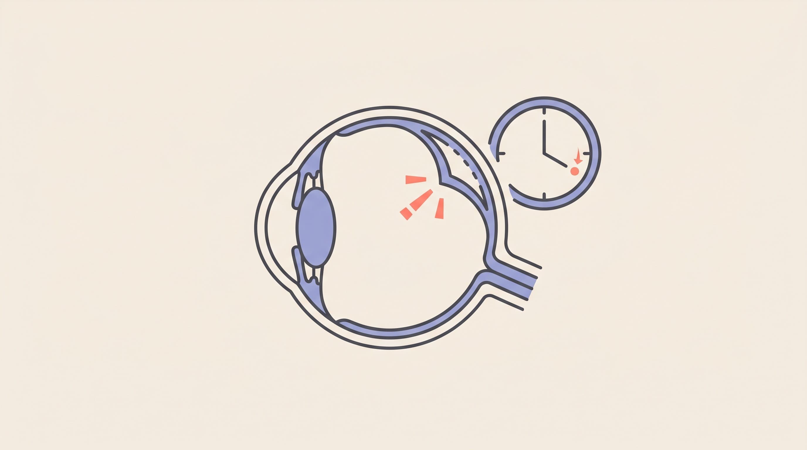
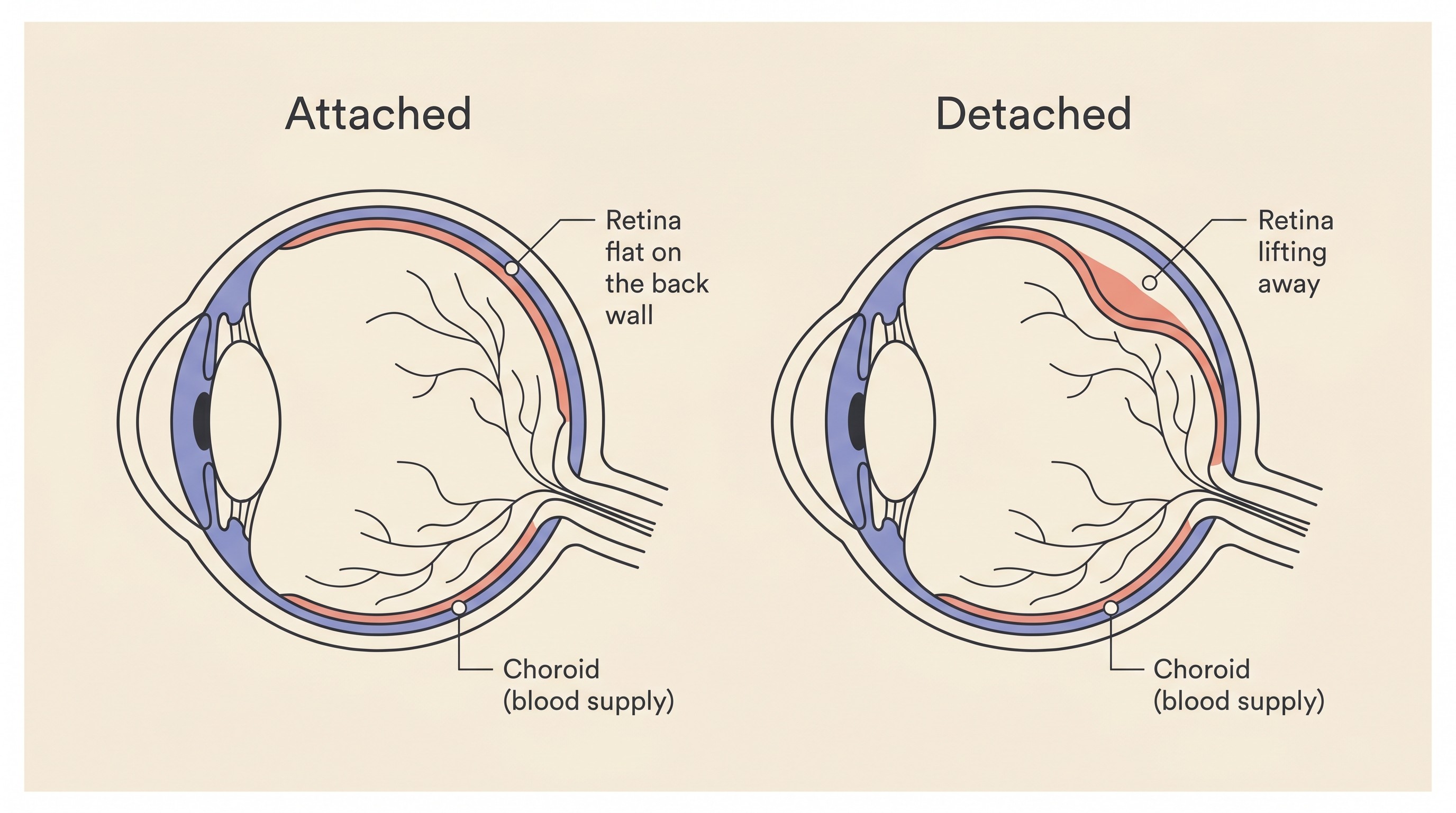
The retina is the thin, light-sensitive layer lining the inside back of the eye, the film at the back of the camera. Its cells, the rods and cones, turn incoming light into the electrical signals that travel down the optic nerve to the brain (StatPearls, 2026). It normally lies flat against the wall of the eye, resting on a blood-vessel-rich layer beneath it called the choroid, which delivers the bulk of its oxygen and nutrients (Cleveland Clinic, 2024).
A detachment is that layer lifting away from the wall. When it lifts, it can't do its job, so the eye stops sending a clear picture to the brain. A vet looking in sees the lifted retina float forward into the eye as a thin grey veil with visible blood vessels, the affected pupil usually wide and slow to react, and if the whole retina has come away, that eye will be blind (Plummer, 2016a).
Here's the part that makes the clock tick. A retina lifted off the wall is also lifted away from that blood supply, and its light-sensing cells are some of the hungriest in the body. Cut off, they start to die, and the longer they're stranded, the more are lost. That is why this is an emergency and not a wait-and-see: the damage is time-dependent, and some becomes permanent if the retina isn't put back against its blood supply quickly.
Three kinds, and why the kind decides everything
This is the most useful thing to understand. Retinas come away by one of three mechanisms, and which one it is largely decides what can be done (Thomasy, 2024; Smith, n.d.).
The common one, by a wide margin, is exudative detachment: fluid builds up underneath the retina and floats it off the wall, like water getting under wallpaper. It happens when the barrier that normally keeps the back-of-eye vessels watertight breaks down and fluid leaks into the space beneath the retina, and in dogs and cats most detachments are this fluid type (Thomasy, 2024; Smith, n.d.). A large pool of fluid down there points to widespread trouble in the choroid, the kind you get with inflammation or very high blood pressure (Smith, n.d.). This is the type those causes produce, and the type that can sometimes settle back down when you fix the underlying problem. That's where the hope in this story lives.
The second is rhegmatogenous detachment, a mouthful that simply means "from a tear or hole." Here the retina is already thinned or frail, from age or degeneration, and a tear lets fluid track behind it (Smith, n.d.). It's less common than the fluid type, and turns up more often in dogs than cats (Thomasy, 2024; Smith, n.d.). A torn retina generally needs an operation, whereas a fluid detachment is usually managed by treating its cause and can sometimes reattach. The third, tractional detachment, something physically pulling the retina off the wall, is the least common of the three (Thomasy, 2024; Smith, n.d.). If you remember nothing else here: the fluid type is the common one, it's the one high blood pressure causes, and it's the one with a real chance of going back down if you move fast.
What causes it
Set out plainly, the causes are a short list (Plummer, 2016a; Plummer, 2016b):
- High blood pressure, especially in cats. The headline cause, and the one most worth acting on fast.
- Inflammation inside the eye (uveitis), often driven by infection or whole-body disease.
- Trauma, a blow to the eye or head.
- Inherited and congenital problems, such as collie eye anomaly, covered in Other Inherited Eye Conditions.
- Eye or body tumours.
- Occasionally after cataract surgery, as a rare complication.
Each is linked where a sibling guide covers it in depth. Here, I want to dwell on the big one.
Why high blood pressure is the headline, and why cats matter most
High blood pressure earns the headline because it's both the commonest cause and the most treatable. The mechanism is simple once you picture it: when the pressure runs too high, fluid is forced out of the vessels at the back of the eye and pools under the retina, peeling it off (Plummer, 2016b). It's the fluid type, which is why prompt treatment can sometimes reverse it.
In cats, this is the version to keep at the front of your mind. The eye is where high blood pressure shows up first and most often, and a detached retina is the classic finding (Cornell Feline Health Center, 2023; Carter, 2019). Across both species, exudative detachment is the most commonly observed sign of blood-pressure damage to the eye, and a sudden, painless loss of sight in both eyes is often the first thing an owner notices (Acierno et al., 2018). In one classic series of 69 hypertensive cats, more than two thirds, 68%, were referred specifically because they had gone blind (Maggio et al., 2000). So an older cat suddenly bumping into things, both pupils wide, the eye normal-looking but unseeing, is more often than not a blood-pressure detachment, which is partly why this is a disease of older, unwell cats rather than a random event.
What's driving that pressure is usually kidney disease or an overactive thyroid in cats, and kidney disease or Cushing's in dogs. The systemic science and the screening message they deserve are owned by High Blood Pressure and the Eyes, and the full cat owner's journey lives in Suddenly Blind Cat? Check the Blood Pressure. If you've got a cat, that second link is the one to follow: a sudden painless detachment in a cat is a check-the-blood-pressure-today problem, not a wait-and-see one.
This isn't a cat-only disease, even so. Dogs get hypertensive detachments too, usually renal or Cushing's-driven, and the torn, congenital and post-surgical types skew towards dogs (Acierno et al., 2018; Thomasy, 2024).
Why it's an emergency, and what can be saved
Speed genuinely changes the outcome. Once a detachment is found, prompt treatment can limit the retinal damage and help vision come back, whereas a retina left detached simply degenerates (Thomasy, 2024). For the common hypertensive detachments, the lever is fast blood-pressure control: bring the pressure down promptly and the retina may lie back down and some sight return (Plummer, 2016a). The window is short. Recovery is best when the detachment has been present for less than about a week (Carter, 2019), and after that the odds fall away. That's exactly what the Eye-emergency triage is built to flag, and the eye-emergency red-flags checklist will settle any doubt.
Now the honest answer to the question you came for, hope and limit in the same breath.
The hope is real. A fluid detachment, especially a hypertensive one caught early, can reattach when the underlying problem is treated, and some sight can come back as the fluid clears (Thomasy, 2024; Plummer, 2016a). In that same series of cats, amlodipine improved the eye signs in 18 of 26 (Maggio et al., 2000). I've seen cats go from blind to navigating again on a daily tablet. Anyone who tells you a detached retina is always hopeless is selling your pet short.
The limit is equally real. The overall outlook for getting vision back is guarded to poor (Plummer, 2016a). If the detachment is chronic, meaning it's been down a while, or came with heavy inflammation, the return of sight is unlikely. And here's the subtle part: even a retina that physically reattaches may not see well, because the cells can degenerate in the meantime, so degeneration is a common sequel even when the layer goes back into place (Plummer, 2016a). The both-species consensus lands in the same spot: effective treatment can reattach the retina, but full vision returns in only a minority, and fixing the pressure doesn't always fix the eye (Acierno et al., 2018).
So the most useful sentence I can give you is this: getting seen fast is about finding out which kind of detachment this is and grabbing the window while it's open, not a promise of a cure. Speed buys the chance, it doesn't guarantee the result. Contrast that with SARDS, which also causes sudden, painless blindness in a normal-looking eye in dogs, but where the retina hasn't detached and so there is nothing to reattach: a detachment can sometimes be treated, SARDS cannot. That's exactly why getting seen fast matters, so each of those two very different stories gets the right response.
What the vet will do, and is surgery an option?
Walking in knowing the plan takes some of the fear out of it. Because high blood pressure is the leading cause, the appointment has a predictable shape. The vet looks into the back of the eye to see the retina directly, and if a detachment or a bleed is found, measures the blood pressure (Plummer, 2016a; Plummer, 2016b). Expect baseline tests too, a blood count, a biochemistry panel and a urine sample to find the underlying disease, and an ultrasound scan if the retina can't be seen behind a bleed or a cataract (Plummer, 2016a; Plummer, 2016b). If high blood pressure is confirmed, the common feline treatment is reassuringly simple: a daily tablet called amlodipine, which lowers the pressure substantially, by 40 to 70 mmHg, and can do it in as little as a week (Carter, 2019), alongside treating whatever is driving it (Cornell Feline Health Center, 2023). The feline workup in full belongs to Suddenly Blind Cat? Check the Blood Pressure.
Most owners picture a "reattachment operation," so let me be straight about where that fits. For the common fluid detachments, the real "surgery" is treating the cause fast: they aren't operated on, they're managed medically, and that's what gives the retina its chance (Thomasy, 2024; Plummer, 2016a). Formal surgical reattachment does exist and has genuinely improved, with published success rates of 72% to 92%, but it's reserved chiefly for the torn detachments and is rare and highly specialised, with very few experienced veterinary retinal surgeons in the world and not every case suitable (Dixon, 2024). So the kind answer to "can it be operated on?" is that for the commonest cause, the operation that matters is treating the blood pressure fast, and that's something your own vet can start today.
When sight doesn't come back
Sometimes, despite everything done right and fast, the sight doesn't return. If that's where you are, there genuinely is a way through. Pets adapt to blindness far better than their owners dare hope, especially once the dust settles. They navigate by a detailed memory map of home, topped up by scent, sound and, in cats, those clever whiskers, and most find their feet within weeks to a few months. The most helpful thing you can do early is keep their world predictable: don't move the furniture, keep food, water and the litter tray exactly where they were, and speak to your pet before you touch them so you never startle them. That whole arc lives in Your Newly Blind Pet: The First 30 Days, with a first-30-days toolkit to match.
But that's the next chapter, and a better one than it sounds from where you're standing. Tonight, if your pet has suddenly lost their sight, your chapter has one action in it. Be seen today, because getting there fast is the only thing that keeps the hopeful door open. Not sure it's urgent? The Eye-emergency triage will tell you in a minute, and for a cat with wide pupils bumping about, you already have your answer.
References
- Acierno, M. J., Brown, S., Coleman, A. E., Jepson, R. E., Papich, M., Stepien, R. L., & Syme, H. M. (2018). ACVIM consensus statement: Guidelines for the identification, evaluation, and management of systemic hypertension in dogs and cats. Journal of Veterinary Internal Medicine, 32(6), 1803-1822.
- Carter, J. (2019). Hypertensive ocular disease in cats: a guide to fundic lesions to facilitate early diagnosis. Journal of Feline Medicine and Surgery, 21(1), 35-45.
- Cleveland Clinic. (2024). Choroid of the eye: what it is, anatomy and function.
- Cornell Feline Health Center. (2023). Hypertension. Cornell University College of Veterinary Medicine.
- Dixon, C. (2024). Canine retinal reattachment surgery: restoring vision. Vet Times, 16 April 2024.
- Maggio, F., DeFrancesco, T. C., Atkins, C. E., Pizzirani, S., Gilger, B. C., & Davidson, M. G. (2000). Ocular lesions associated with systemic hypertension in cats: 69 cases (1985-1998). Journal of the American Veterinary Medical Association, 217(5), 695-702.
- Plummer, C. E. (2016a). Retinal detachment. Clinician's Brief, January 2016.
- Plummer, C. E. (2016b). Diagnosing acute blindness in dogs. Today's Veterinary Practice, 18 October 2016.
- Smith, P. J. (n.d.). Canine retinal detachment (Chapter 76). Veterian Key.
- StatPearls. (2026). Neuroanatomy, retina. NCBI Bookshelf.
- Thomasy, S. M. (2024). Retinal detachment in small animals. MSD Veterinary Manual (Professional).
Keep track of how your pet is doing
The owners who cope best are the ones who notice changes early. A simple health log shows you what is working, and what is not, before the next vet visit.
Start tracking, freeYou're not doing this alone
Compare treatment journeys and talk to owners managing vision & eye health. Free to join.
Join PetsLikeMine