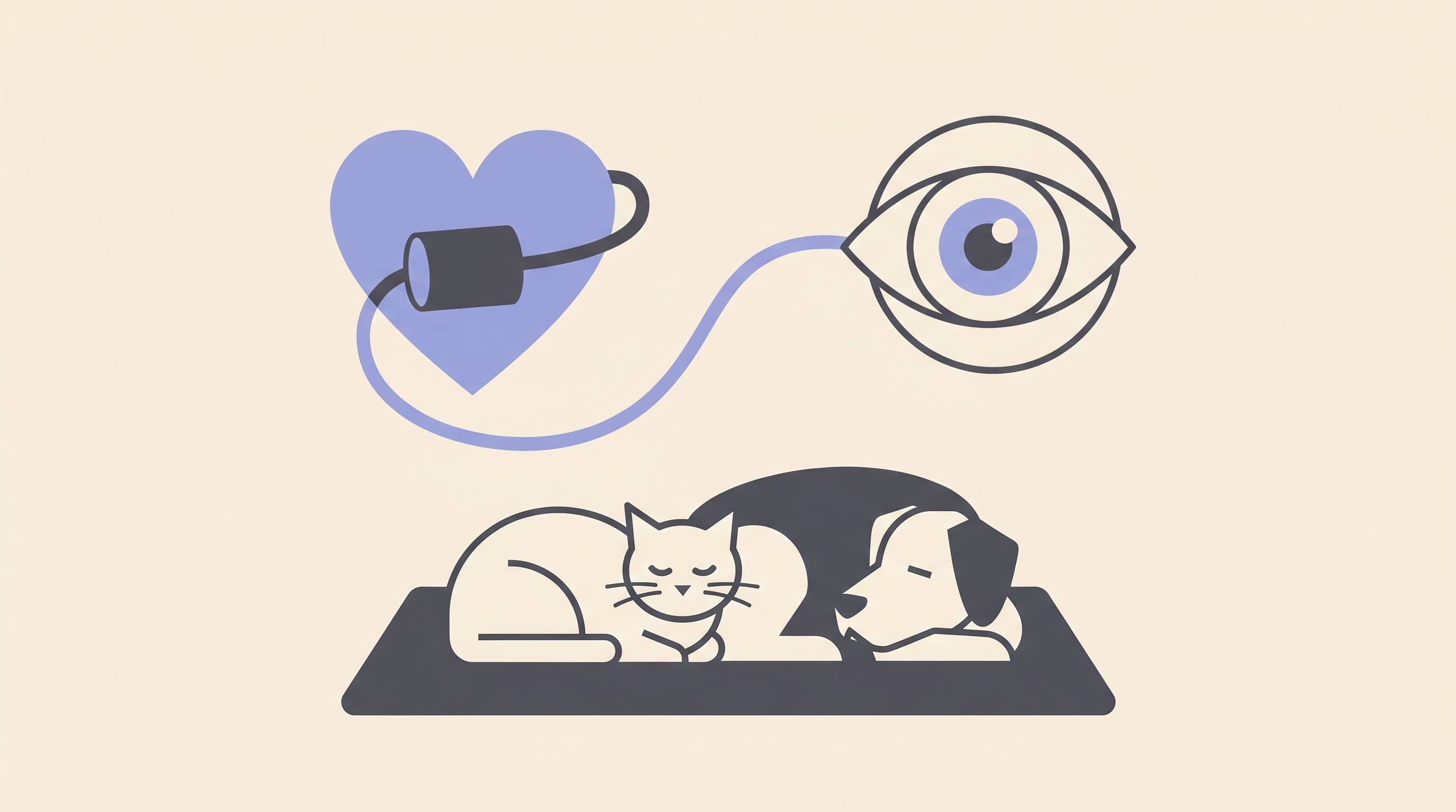
High Blood Pressure and Your Pet's Eyes: The Link Owners Miss
Claire Greenway
BVM&S MRCVS
Most of us think of high blood pressure as a human problem, the sort of thing a doctor mentions at a midlife check-up. So when I tell an owner that their cat's sudden blindness, or the red haze that's appeared in their dog's eye overnight, is down to blood pressure, the reaction is almost always the same: a pause, then "pets get that?" They do. And here is the part that catches everyone out. In animals, the eye is often the very first place high blood pressure shows itself, and by the time it does, sight is already on the line.
This article is about that link, plainly: why high blood pressure damages the back of the eye, what drives the pressure up (and how that differs a little between cats and dogs), and, most importantly, the genuinely preventable bit. Unlike a lot of what I write about, this is a problem we can often catch before the eye is lost, if the right pets get a simple, painless check at the right time. If your pet has just gone suddenly blind or has visible blood in an eye right now, this is a same-day emergency: ring your vet, and our Eye-emergency triage will help you judge how fast. For everyone else, let me start with why the eye is such a giveaway.
Why the eye is the first to show it
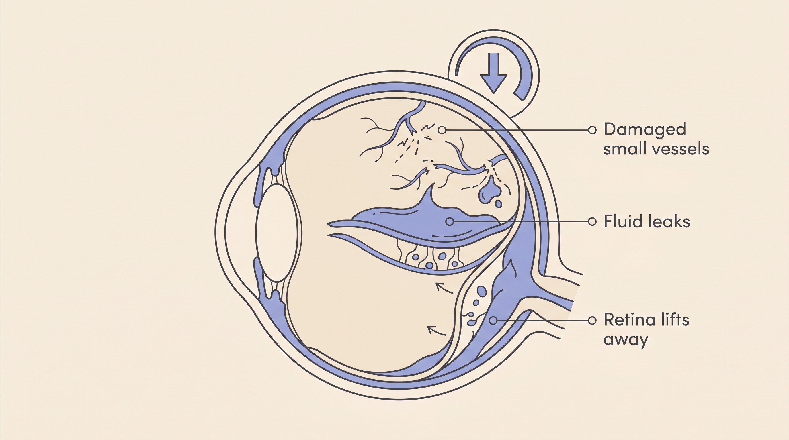
The back of the eye holds one of the busiest networks of small blood vessels anywhere in the body, and that's exactly why it's vulnerable. Sustained high blood pressure injures the walls of those tiny arterioles, and across the body it tends to pick on the organs with the richest arteriolar supply: the eyes, the kidneys, the heart and the brain (Taylor et al., 2017). The difference with the eye is that it's the one you can sometimes see into. The others fail quietly.
What happens inside the eye follows a fairly predictable course. The vessels first clamp down to try to autoregulate, then their walls become occluded and finally start to leak (Brown, 2022). Fluid and blood seep out from the layer of vessels behind the retina (the choroid) and pool underneath the light-sensing retina, lifting it away from the back of the eye like wallpaper off a damp wall. In cats this leak is the heart of the problem: the subretinal fluid originates from the choroidal vessels (the choriocapillaris), and as it builds it can lift the retina into a serous detachment (Carter, 2019). A fluid-driven (exudative) retinal detachment is in fact the most common ocular finding of systemic hypertension (Acierno et al., 2018). Alongside it, vets commonly see bleeds within the retina, blood in the front of the eye (hyphema), and retinal vessels gone tortuous, taking on a corkscrew shape (LeBlanc et al., 2011; Brown, 2022). How a retina detaches, and what can be salvaged, belongs to retinal detachment; I'll stay on the blood-pressure thread here.
To an owner, none of that is visible from the sofa. What you notice is the consequence: a pet that has suddenly started bumping into things, often with pupils that stay wide and don't shrink in bright light, and sometimes a red tinge or a pool of blood inside the eye. The vision loss itself comes from that cocktail: intraocular bleeding, swelling under the retina, the detachment, and occasionally a secondary glaucoma if blood blocks the eye's drainage (Brown, 2022). Which is why the single most useful rule in the whole space applies here: a cat that suddenly can't see needs its blood pressure checked the same day, not next week. The owner side of that story, the ring-the-vet-now headline and what to expect, lives in cat sudden blindness and blood pressure.
And it really is a first sign more often than a late one. In dogs seen at a referral hospital for confirmed high blood pressure, "sixty-two percent (26/42) of hypertensive dogs had [at least] 1 type of ocular lesion identified" (LeBlanc et al., 2011), and in cats, ocular problems are the most common form of target-organ damage seen at all (Cornell Feline Health Center, 2023). The reason the eye matters so much for catching this early is that the damage there is, in one specialist's words, "the most easily identifiable form" of that organ damage, and often the very reason the animal is brought in (Carter, 2019). Across both species, the eye changes are "often an initial clinical sign" of the whole problem (Brown, 2022). I'd add one honest caveat so you don't over-read it: a normal-looking eye does not rule high blood pressure out. In that canine series, eye lesions flagged hypertension only about six times in ten, so a clear eye is reassuring but not a guarantee (LeBlanc et al., 2011). The eye, in effect, is a smoke alarm for a fire happening elsewhere in the body: when it sounds, take it seriously, but don't assume silence means there's no fire. Which brings us to the question that matters for prevention: what lit the fire?
What's driving the pressure up: cats
Here is the crux of why this article exists, and why it links out to so many others. High blood pressure in pets is almost never a disease in its own right. It is secondary in at least eight cases out of ten, a symptom of something else, and the usual culprits differ a little between cats and dogs (Acierno et al., 2018).
In cats, the two names that come up again and again are chronic kidney disease and an overactive thyroid (hyperthyroidism). Kidney disease leads by a distance: at least 60% of cats diagnosed with high blood pressure also have signs of chronic kidney disease (Cornell Feline Health Center, 2023), and it's the most frequently reported cause of hypertension across both species (Acierno et al., 2018). An overactive thyroid is the other big one, with around 20% of hyperthyroid cats turning out to be hypertensive too (Cornell Feline Health Center, 2023). So when I find a hypertensive bleed in an older cat's eye, I'm not finished, I'm just getting started: that eye is telling me to look hard at the kidneys and the thyroid. How kidney disease is staged and managed, and how an overactive thyroid is treated, sits with the spaces that own them: our CKD space and the Hormone Health home. My job here is to draw the line between them and the eye.
What's driving the pressure up: dogs
Dogs are not an afterthought here, even though the feline picture is the better known one. The drivers are similar but the order shifts. Chronic kidney disease is again a leading cause, but the name that gives the canine version its character is Cushing's disease, the syndrome of overactive adrenal glands (hyperadrenocorticism). The association is strong: in a study of dogs with naturally occurring Cushing's, more than four in five were hypertensive, with high blood pressure found in 82% of them (García San José et al., 2020). Behind these sit other causes the consensus panel lists, including diabetes and rarer hormone-producing tumours (Acierno et al., 2018). In the landmark canine eye series, the underlying diseases were exactly this cast: kidney disease most often (11 of the dogs), then diabetes (5), then Cushing's (3) and the occasional adrenal tumour (LeBlanc et al., 2011).
So a hypertensive eye in a dog is a prompt to investigate the kidneys and the adrenal glands, not the end of the story. Cushing's in dogs (and the thyroid in cats) is covered in the Hormone Health home; kidney disease has the CKD space. One distinction trips dog owners up: a dog that goes suddenly and completely blind with a normal-looking, non-painful eye may not have a blood-pressure problem at all but a separate condition called SARDS, which has no treatment. Confusingly, SARDS dogs are often thirstier and hungrier beforehand, which overlaps with Cushing's signs, but it's a different thing from a hypertensive bleed, and the distinction matters because hypertensive blindness is treatable if caught fast whereas SARDS is not. SARDS draws out that contrast in full.
The preventable part, and why I keep banging on about it
This is the bit I most want to land, because it's the rare place in eye medicine where an owner genuinely holds a lever. The pressure can be found and brought down before the eye is lost, but only if the at-risk pet actually gets it measured, and far too many never do.
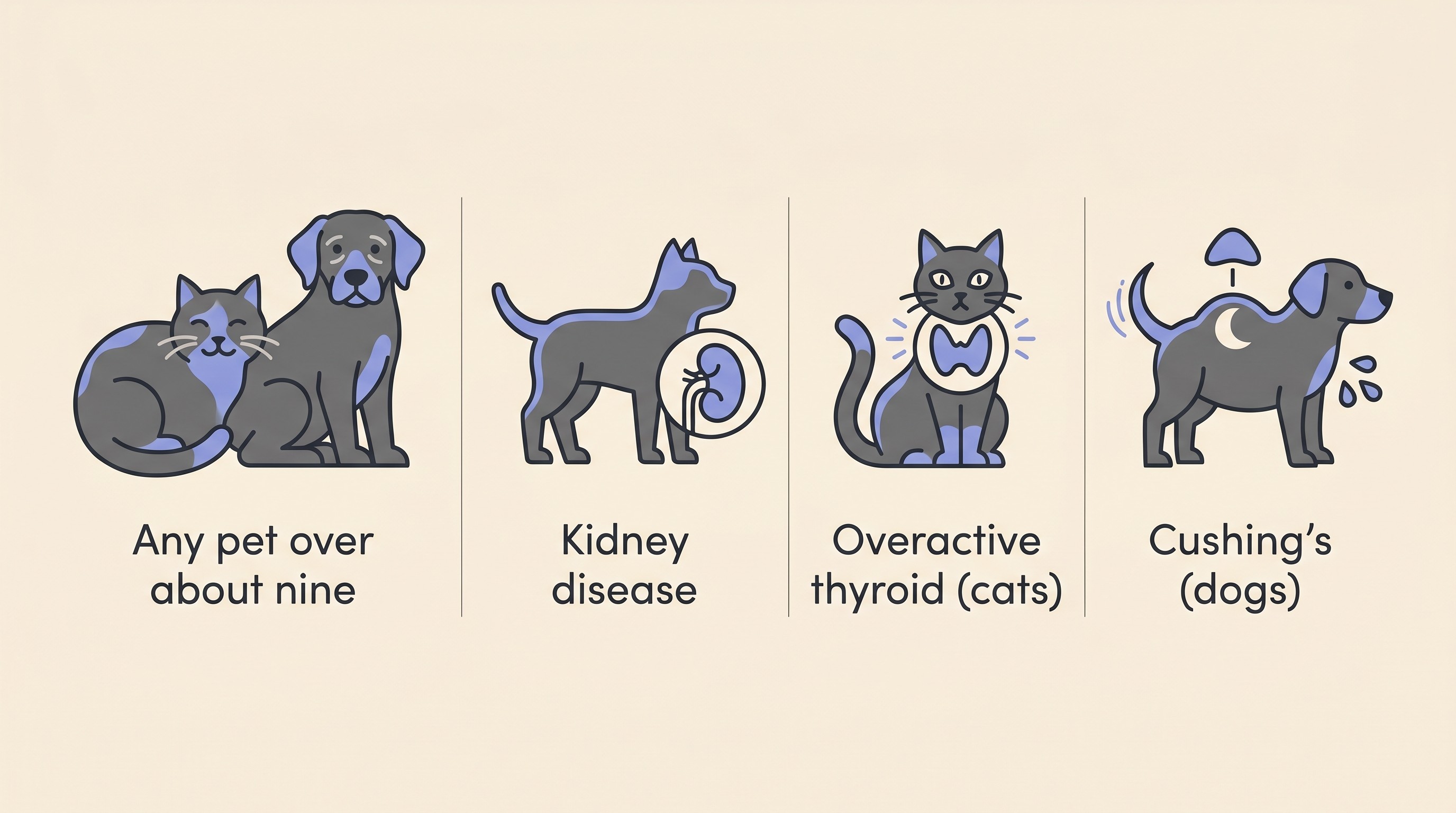
The good news is that the advice is targeted, not a counsel of perfection. The consensus guidance is that it's reasonable to start annual blood-pressure screening in cats and dogs from around nine years of age, and the panel explicitly does not call for screening every pet of every age (Acierno et al., 2018). On top of that age threshold, the pets who really need it are the ones with a disease that drives pressure up. For cats, the guidance is firm: those with any underlying condition, especially kidney disease or an overactive thyroid, should have their blood pressure screened regularly (Cornell Feline Health Center, 2023). The feline gold-standard guideline puts numbers on the cadence: at least every 6 to 12 months in geriatric cats, and at least every 3 to 6 months in cats with kidney disease or an overactive thyroid (Taylor et al., 2017). The same logic carries to dogs with kidney disease or Cushing's.
So the call to action is simple, and you can act on it at your next appointment. If your pet has kidney disease, an overactive thyroid, Cushing's, or is simply getting on in years, ask whether their blood pressure has been checked recently.
The check itself is undramatic: an inflatable cuff, usually on a paw or the tail, a couple of minutes, no needles, no fuss. It's the kind of quiet, unglamorous test that saves eyes precisely because it catches the problem while there's still something to save. And even setting the eye aside, the same high pressure is quietly working on the kidneys, the heart and the brain too (Taylor et al., 2017). The eye is just the one that lets us catch it in the act.
The honest hope, and the treatment
I want to be straight about outcomes, because this is one of the few sudden blindnesses where speed genuinely changes the ending, and I never want to oversell or undersell it.
If high blood pressure is caught and brought down quickly, the fluid and blood lifting the retina can gradually clear, the retina can settle back into place, and some, occasionally all, of the lost sight can return, though that recovery isn't instant and can take weeks to months (Veterinary Vision Center, 2023). Timing is everything: recovery of a detached retina is best when it has been lifted for less than a week, which is exactly why "today" is not me being dramatic (Carter, 2019). But here's the honest other half: by the time many pets are presented, the damage is already done and the loss is permanent (Cornell Feline Health Center, 2023). Both things are true at once. Sudden onset does not mean salvageable, and the difference between the eye that recovers and the eye that doesn't is very often measured in days. That tension is the entire argument for screening: we can sometimes reverse this one, which is exactly why we don't want to be meeting it for the first time after the retina has already let go. The detailed feline recovery figures, how often sight comes back and how fully, live with cat sudden blindness and blood pressure rather than being restated here.
Treatment runs along two tracks at once: bring the pressure down, and treat the disease underneath it. The tablet differs by species, and it matters to get right. In cats, the first-line medication is amlodipine, which has the best-established track record in the species (Acierno et al., 2018). In dogs, the usual starting choice is an ACE inhibitor such as benazepril or enalapril, with amlodipine added when the pressure is very high or an ACE inhibitor alone isn't enough, and the aim either way is to steer the systolic pressure back toward normal (Acierno et al., 2018). I'll leave the doses to your own vet, who's measuring the response, and the underlying kidney, thyroid or adrenal disease to the spaces that own those subjects. The point to hold is that lowering the pressure and treating the cause go hand in hand, and the eye improves when both are happening.
If sight comes back, wonderful. If it doesn't, this is not the end of a good life, and I'd never want an owner to think otherwise. Pets adapt remarkably well to blindness, especially older cats, who lean heavily on smell, hearing and their whiskers and often surprise their owners with how quickly they find their feet. Helping a newly blind pet through the first weeks, the small changes that matter, the golden rule of not moving the furniture, all sit in the living-with cluster, and newly blind: the first 30 days is the place to start.
A hypertensive eye bleed is a genuine emergency in the moment. But it's also, more than almost any other cause of sudden blindness, a problem we can sometimes get ahead of. So if you take one thing from this page, let it be the cuff: for an older pet, or a pet with kidney disease, an overactive thyroid or Cushing's, ask for the check before the eye ever has the chance to make the point for you. If you'd like a single sheet to take to that appointment, our feline blindness and blood pressure download lays the whole thread out on one page.
References
- Acierno, M. J., Brown, S., Coleman, A. E., Jepson, R. E., Papich, M. G., Stepien, R. L., & Syme, H. M. (2018). ACVIM consensus statement: Guidelines for the identification, evaluation, and management of systemic hypertension in dogs and cats. Journal of Veterinary Internal Medicine, 32(6), 1803-1822.
- Brown, M. H. (2022). Top 5 hypertension-related eye abnormalities in dogs & cats. Clinician's Brief, June 2022.
- Carter, J. (2019). Hypertensive ocular disease in cats: a guide to fundic lesions to facilitate early diagnosis. Journal of Feline Medicine and Surgery, 21(1), 35-45.
- Cornell Feline Health Center. (2023). Hypertension. Cornell University College of Veterinary Medicine.
- García San José, P., Arenas Bermejo, C., Clares Moral, I., Cuesta Alvaro, P., & Pérez Alenza, M. D. (2020). Prevalence and risk factors associated with systemic hypertension in dogs with spontaneous hyperadrenocorticism. Journal of Veterinary Internal Medicine, 34(5), 1768-1778.
- LeBlanc, N. L., Stepien, R. L., & Bentley, E. (2011). Ocular lesions associated with systemic hypertension in dogs: 65 cases (2005-2007). Journal of the American Veterinary Medical Association, 238(7), 915-921.
- Taylor, S. S., Sparkes, A. H., Briscoe, K., Carter, J., Sala, S. C., Jepson, R. E., Reynolds, B. S., & Scansen, B. A. (2017). ISFM consensus guidelines on the diagnosis and management of hypertension in cats. Journal of Feline Medicine and Surgery, 19(3), 288-303.
- Veterinary Vision Center. (2023). Hypertensive retinopathy in dogs and cats.
Keep track of how your pet is doing
The owners who cope best are the ones who notice changes early. A simple health log shows you what is working, and what is not, before the next vet visit.
Start tracking, freeYou're not doing this alone
Compare treatment journeys and talk to owners managing vision & eye health. Free to join.
Join PetsLikeMine