How Your Pet's Heart Works, and What Can Go Wrong
Dr. Alastair Greenway
MRCVS
If you have just heard the word "heart" from your vet, your mind is probably racing ahead to the worst version of the story. Take a breath. Before any diagnosis makes sense, and before you can tell which articles in this space are actually about your pet, it helps to understand what the heart is doing in the first place, and the small number of ways it tends to go wrong. There are really only three common patterns, and almost everything that follows in cats and dogs is a variation on one of them. This article is the plain-language map of the territory. It will not diagnose your pet, but it will help you understand what your vet has already said and point you to the right next read.
The heart is just a pump, with four rooms
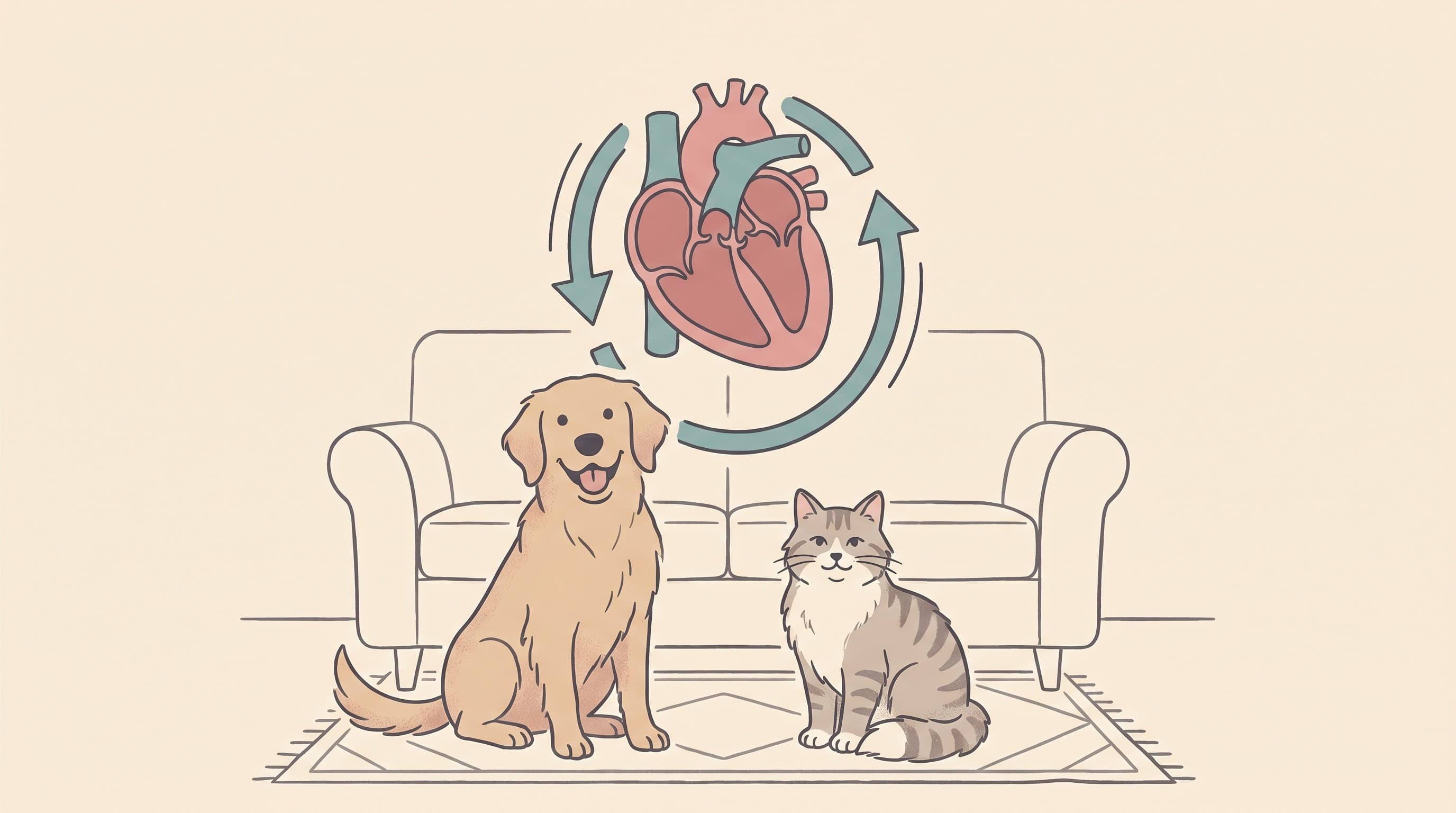
Strip away the anatomy and the heart is a muscular pump with one job: to keep blood moving in a loop, around and around, for the whole of your pet's life. It does this without a single rest, beating roughly 70 to 140 times a minute in a dog and 140 to 220 times a minute in a cat, every minute, for years on end. When you understand that it is fundamentally a pump, the things that go wrong start to make intuitive sense, because a pump can only fail in a few ways.
The heart has four chambers, arranged as two pumps side by side. The two upper chambers, the atria, are the receiving rooms: blood arrives here from the body and the lungs. The two lower chambers, the ventricles, are the powerful pushing rooms that do the heavy work of sending blood back out. Between each upper and lower chamber sits a one-way valve, a pair of thin flaps that snap shut after every beat so blood cannot slosh backwards. That click of the valves closing is, in fact, most of the "lub-dub" you hear through a stethoscope.
The loop itself has two halves. The right side of the heart collects oxygen-poor blood returning from the body and pumps it the short distance to the lungs, where it picks up oxygen. The freshly oxygenated blood comes back to the left side, which is the muscular one, and the left ventricle pumps it out under pressure to the whole body. Then the cycle repeats. Hold on to that picture of two pumps, four rooms and a set of one-way valves, because every problem below is simply one of those parts not doing its job.

The three ways a heart goes wrong
For all the frightening medical names, the common heart diseases of dogs and cats fall into three buckets. It is genuinely worth learning them, because once you know which bucket your pet sits in, the rest of this space falls into place.
A leaky valve. This is by far the most common heart problem in dogs. Over the years one of those one-way valves, usually the mitral valve on the powerful left side, thickens and degenerates so its flaps no longer seal cleanly. Every time the heart beats, a little blood squirts backwards through the gap instead of all going forwards. That backward jet of blood is turbulent, and turbulence is noisy, which is why a leaky valve is the classic cause of a heart murmur in a dog (Atkins et al., 2009). The disease has several names that mean broadly the same thing: myxomatous mitral valve disease, degenerative valve disease, or endocardiosis. It is the bread-and-butter heart disease of the older small-breed dog, and Cavalier King Charles Spaniels are especially prone to it (Borgarelli and Buchanan, 2012). If your dog has been told they have a "leaky valve" or "mitral valve disease", start with the mitral valve disease explainer.
A thick heart. This is the cat's disease. Here the muscular wall of the left ventricle grows abnormally thick, so the chamber becomes stiff and cannot relax and fill properly between beats. The pump still squeezes, often perfectly well, but it cannot accept blood easily, and a heart that cannot fill cannot push out a full load. This is hypertrophic cardiomyopathy, or HCM, and it is the most common heart disease in cats by a wide margin: screening studies of apparently healthy cats have repeatedly found it in around one in seven (Paige et al., 2009; Payne et al., 2015). If "thickened heart muscle" or "HCM" is what you have been told, the feline HCM explainer is your starting point.
A weak heart. In this pattern the heart muscle itself becomes thin, baggy and weak, so the ventricle stretches out and loses its squeeze. A weak, dilated pump cannot move blood forwards with enough force. This is dilated cardiomyopathy, or DCM, and it is primarily a disease of large and giant-breed dogs, with Dobermanns and several other breeds carrying a genuine genetic predisposition (Wess et al., 2017). It is much rarer in cats than it once was, for a reason that is one of veterinary medicine's great success stories: classic feline DCM turned out to be caused by a deficiency of the amino acid taurine in cat food, and once pet food manufacturers corrected this in the late 1980s, the disease all but vanished (Pion et al., 1987). In dogs there is now a separate, actively researched concern about certain grain-free diets and DCM, which has its own dedicated read: the grain-free question.
So: a leaky valve in dogs, a thick heart in cats, and a weak heart mostly in big dogs. Three pictures, three buckets. Almost every owner reading this space has a pet in one of them.
Why "backward pressure" is the thing to understand
Here is the single most useful idea in this whole article, because it explains the symptom that frightens owners most: the breathing.
Whichever of the three problems a pet has, they share a common downstream consequence. A leaky valve, a stiff thick heart and a weak baggy heart all, in their own way, make it harder for blood to move forwards through the pump. And blood that cannot move forwards efficiently has to go somewhere, so it backs up, like traffic queueing behind a bottleneck. Pressure rises behind the struggling chamber.
When that pressure rises behind the left side of the heart, it pushes back into the lungs, because the lungs sit just upstream of the left side. Eventually the pressure forces watery fluid out of the tiny blood vessels in the lungs and into the air spaces themselves. This is pulmonary oedema: fluid where air should be. A pet with fluid in their lungs cannot get oxygen in properly, so they breathe faster and harder, and that is the moment a quiet heart problem becomes a visible, urgent illness. This is what the word "congestion" in congestive heart failure actually refers to: the congestion of fluid that backs up behind a failing heart.
The detail varies a little by species. In dogs the fluid almost always ends up inside the lung tissue, causing that fast, laboured breathing and often a cough. In cats it may collect inside the lungs too, but cats are also prone to fluid pooling in the chest cavity around the outside of the lungs, a pleural effusion, which leaves them breathing in a shallow, restricted way. Either way, the common thread is breathing that has changed.
This is exactly why the most powerful thing you can do at home costs nothing and needs no equipment: counting how fast your pet breathes while they are fast asleep. A resting breathing rate that starts climbing is often the very first sign that fluid is beginning to build, frequently before your pet seems unwell to you, and it is the best early-warning number you have (Ohad et al., 2013; Porciello et al., 2016). We treat it as important enough to deserve its own full guide: the resting respiratory rate guide shows you exactly how to count it and what is normal, and the breathing-rate tracker turns it into a daily habit with a chart you can show your vet. If your pet is breathing rapidly or with effort right now, that is not a number to log, it is a reason to act: the heart failure emergency signs article walks through what counts as a crisis and what to do.
Dogs and cats are different patients
It is worth being honest about how differently these two species behave, because it changes what you should be watching for.
Dogs tend to advertise. A dog with developing heart disease will often cough, tire on walks, or breathe faster, and because most canine heart disease is a slowly worsening leaky valve, there is usually a long, quiet window of years between the first murmur and any real trouble. That window is genuinely good news, because it gives you time to monitor and, in some cases, to start treatment that delays the onset of heart failure: in dogs with significant preclinical mitral valve disease, beginning the medication pimobendan before any symptoms appear was shown to meaningfully extend the symptom-free period (Boswood et al., 2016). Not every dog with a murmur is anywhere near that stage, which is why the murmur itself is a prompt to investigate, not a diagnosis. The what a murmur means article unpacks that properly.
Cats, by contrast, are infuriatingly good at hiding it. A cat is a small, sedentary ambush predator, and a cat that feels unwell simply does less: it sleeps more, plays less, and tucks itself away, all of which look like ordinary cat behaviour. Many cats with significant HCM have no murmur at all, so a clean listen through the stethoscope is reassuring but never a guarantee (Payne et al., 2015). The unhappy result is that in a meaningful number of cats the very first sign anyone sees is a sudden crisis: a cat that is abruptly breathing hard, or one whose back legs become suddenly painful and paralysed because a blood clot has formed in the enlarged heart and lodged in the arteries to the hind limbs. This clot complication, feline arterial thromboembolism, is one of the reasons feline heart disease is treated with such respect, and it is covered in the cat articles in this space. The practical message is simple: with cats, you cannot rely on them to tell you, so the home breathing count and your vet's screening matter even more.
Where to go next
You now have the map, so you can head straight for the part that is actually about your pet. The fastest route is to follow what your vet has already said.
If your dog has a murmur or a leaky valve, read what a murmur means and then the mitral valve disease explainer. If your cat has been told they have a thickened heart or HCM, go to the feline HCM explainer. If a large-breed dog has a weak or enlarged heart, or DCM, start with the grain-free question and the screening guidance for at-risk breeds. If your vet has used the words heart failure or fluid on the lungs, the congestive heart failure explainer explains what that means and what treatment looks like.
And whatever the underlying problem, two things apply to almost everyone here. If your vet wants to run tests to pin down the diagnosis, the tests explained tells you what an x-ray, an echo, an ECG, a blood pressure check and a proBNP blood test each show and what to expect. And from today onwards, start getting to know your pet's resting breathing rate, because that one number, tracked quietly while they sleep, is the thread that runs through this entire space.
Keep track of how your pet is doing
The owners who cope best are the ones who notice changes early. A simple health log shows you what is working, and what is not, before the next vet visit.
Start tracking, freeYou're not doing this alone
Compare treatment journeys and talk to owners managing heart health. Free to join.
Join PetsLikeMine