

Feline HCM Explained: The Thickened Heart, and Why Cats Hide It
Alastair Greenway
BVM&S MRCVS
Hypertrophic cardiomyopathy is a mouthful of a diagnosis, and most owners leave the consulting room holding the letters HCM and not much else. Meanwhile the cat it refers to is asleep on the sofa, looking entirely fine. That mismatch, between a name that sounds enormous and an animal that seems perfectly well, runs through the whole of this disease, so it is worth taking slowly. Think of this as the plain-English anchor for feline HCM. We will work through what the thickened heart is, why so many cats have it, why it usually gives no warning, where it can lead, and what you can actually do. The sibling articles dig deeper into each part, and I will point you to them as we go, but the foundations are all here.
What HCM actually is
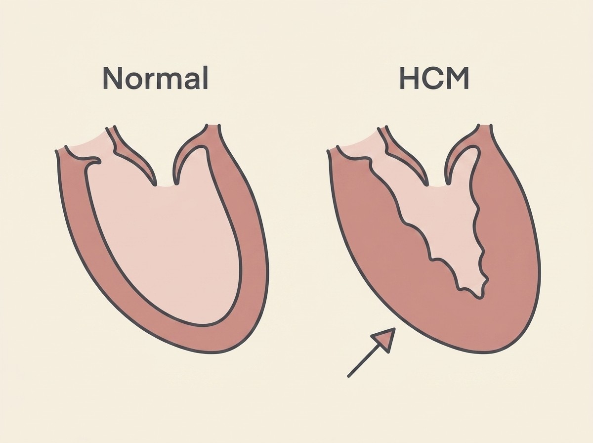
The heart is a four-chambered pump, and the chamber that does most of the hard work is the left ventricle, the muscular lower-left chamber that drives blood out to the body. In hypertrophic cardiomyopathy, the muscular wall of that chamber grows abnormally thick. The name pulls apart neatly: "hypertrophic" means overgrown, "cardio" means heart, "myopathy" means a disease of the muscle. A disease of overgrown heart muscle.
You might expect a thicker, more muscular heart to be a stronger one. It works the other way round. A thickened wall is a stiff wall, and a stiff ventricle cannot relax and stretch open properly between beats. Because it cannot relax, it cannot fill easily, and a chamber that cannot fill cannot send out a full load with each beat (Luis Fuentes et al., 2020). The squeeze, the pumping action, is often perfectly good. It is the filling that fails. That is why HCM is described as a disease of a heart that struggles to relax rather than one that struggles to pump, and that single distinction shapes almost everything about how it behaves.
The most common heart disease in cats
HCM is not a rare or exotic diagnosis. By a wide margin it is the commonest heart disease in cats, and far more widespread than most owners ever realise. When researchers have taken groups of apparently healthy cats, animals with no known heart problem at all, and scanned every one of them, they have repeatedly found that roughly one in seven was quietly carrying cardiomyopathy, the great majority of it HCM (Paige et al., 2009). A large UK study of cats in rehoming centres, the CatScan study, landed in the same place: a prevalence close to 15% in ordinary, healthy-seeming cats (Payne et al., 2015).
Those numbers change how the diagnosis feels. If your cat has HCM, your cat is not some strange unlucky exception. A real slice of the entire cat population is walking around with a thickened heart nobody has spotted. That is sobering, and it is also oddly steadying, because it means a great deal of veterinary research has gone into this exact condition, and there is a well-mapped path through it.
Why it is so often silent
Here is the part of feline HCM every owner needs to take on board: most of the time, it makes no noise at all. Cats are extraordinary at hiding illness, an instinct handed down from ancestors for whom looking weak was dangerous, and HCM slips straight through that instinct. Plenty of cats with a thickened heart look and behave completely normally. They eat, they play, they jump, they purr, and nothing about them says "heart disease".
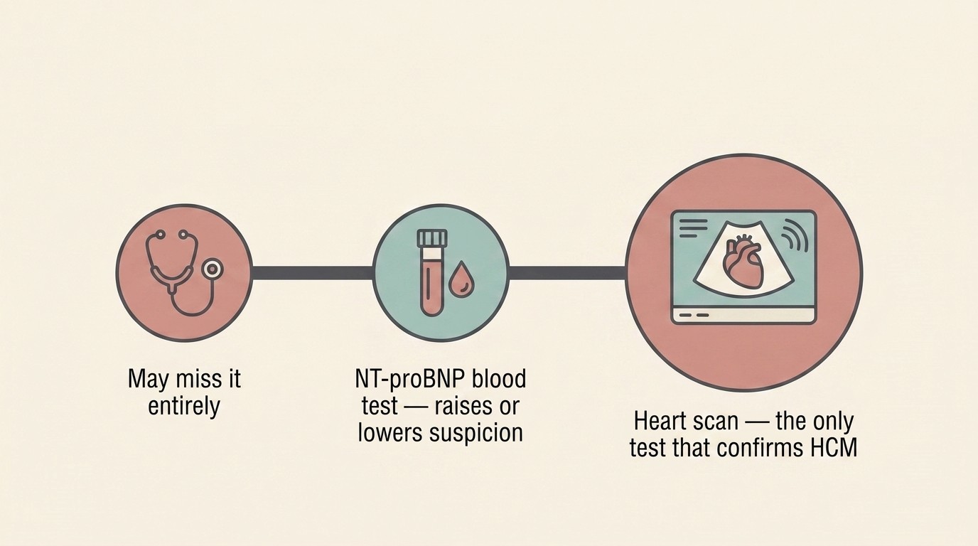
It is tempting to assume a vet will simply hear the problem through a stethoscope, and sometimes that is exactly how it surfaces. A vet may catch a heart murmur, an extra whooshing sound, or a gallop rhythm during a routine check, and that prompts a closer look. But the stethoscope is an unreliable detective here, and it misleads in both directions. Plenty of cats with HCM have no murmur whatsoever, so a clean listen is not a clean bill of heart health. And plenty of cats with a murmur have a perfectly normal heart, because murmurs in cats are often innocent or down to something other than HCM (Paige et al., 2009). A stethoscope can raise a suspicion. It cannot confirm or rule out HCM, which is why so many cases stay hidden, and why screening exists at all.
The three dangers HCM can lead to
A thickened heart matters because of where it can lead. Most cats with mild HCM never travel down these roads, and that is worth holding onto throughout. Still, you deserve to know the three serious events HCM can set off, because spotting them fast is sometimes what matters most.
The first is congestive heart failure. When the stiff ventricle cannot relax, pressure backs up behind it, into the left atrium and then into the blood vessels of the lungs. Eventually fluid is forced out into or around the lungs, and the cat begins to struggle for breath. In cats this tends to show as fast or laboured breathing, or open-mouth breathing, rather than the cough owners often expect. What this looks like at home, and what to do, is covered fully in when a cat with HCM goes into heart failure.
The second is arterial thromboembolism, often called a saddle thrombus. A clot can form in the enlarged, sluggish left atrium, break loose, and lodge where the main artery divides to supply the back legs. The result is sudden, agonising paralysis of one or both hind limbs, with cold, pale pads and obvious distress, and it is sometimes the very first sign that a cat had heart disease at all. It is a true emergency, and because every HCM owner should be able to recognise it in seconds, it has its own dedicated guide: saddle thrombus, the emergency every HCM owner must know.
The third is sudden death. In a smaller number of cats, HCM disturbs the heart's electrical rhythm badly enough to cause collapse and death with little or no warning (Luis Fuentes et al., 2020). This is the rarest of the three and the hardest to predict, and dwelling on it helps no one, but being honest about HCM means naming it.
Hold this alongside the rest: a large international study that followed cats diagnosed with HCM who showed no outward signs found that many stayed completely free of any of these events for years, especially the cats whose hearts were only mildly affected (Fox et al., 2018). The dangers are real, but for most cats they are possibilities to watch for, not certainties to brace against.
How HCM is actually found
Because the stethoscope cannot settle the question, finding HCM properly relies on tests that look past it. Two of them matter.
The first is a heart scan, or echocardiogram, an ultrasound of the heart. This is the only test that truly diagnoses or excludes HCM, because it is the only one that looks directly at the heart wall and measures how thick it actually is (Luis Fuentes et al., 2020). Ideally a cardiologist or an experienced operator performs it. Everything else points towards the scan. The scan is what sees.
The second is a blood test called NT-proBNP, which rises when heart muscle is under strain. There is a quick in-clinic SNAP-type version and a more accurate quantitative laboratory version (Fox et al., 2011). Treat it as a useful nudge rather than a verdict: a raised result is a strong reason to arrange a scan, while a normal in-clinic result does not reliably clear the heart on its own. How the gene tests, the blood tests and the scan fit together, and which question each one really answers, is laid out in screening and genetics.
Predisposed breeds, but any cat
Some breeds carry a known, inherited layer of risk. In Maine Coons, a specific mutation in the MYBPC3 gene, the A31P variant, is associated with HCM, and a different MYBPC3 mutation, the R820W variant, has been described in Ragdolls (Meurs et al., 2005; Meurs et al., 2007). A cheek-swab test can identify carriers of these variants, which is genuinely useful for breeders making pairing decisions.
Two things need saying plainly, though. A negative gene test does not clear a cat of HCM, because the known mutations explain only a fraction of cases, and many cats with classic HCM test negative for everything we can currently look for. And HCM is in no way confined to pedigree cats. It turns up right across the whole cat population, including ordinary domestic shorthairs and moggies with no special breeding at all. The breeds simply add a partly-mappable layer of risk on top of a baseline that every cat shares. If you own a non-pedigree cat, HCM is still firmly on the table.
What you can do
The most powerful tool you have at home costs nothing and takes about thirty seconds. It is counting your cat's resting respiratory rate, the number of breaths taken in a minute while your cat is settled or asleep. A healthy cat at rest usually breathes fewer than around 30 times a minute, and a rate that climbs and stays high is frequently the earliest sign that fluid is starting to gather around the lungs, often a day or two before a cat looks unwell. Because cats hide so much, this quiet number can be the first crack in the disguise. The resting respiratory rate guide explains exactly how to count it, and the breathing rate tracker turns it into an easy nightly habit so a rising trend stands out rather than being lost to a vague sense that "she seems a bit puffed lately".
The other thing within your control is screening at-risk cats. If you have a known predisposed breed, a breeding cat, or a cat with a murmur or an upcoming non-urgent anaesthetic, a heart check before trouble appears is the single most valuable step. A stiff heart copes poorly with the stress and the drugs of an operation, which is why an at-risk cat is often best scanned first; HCM and anaesthesia covers exactly who needs checking and why.
A final, honest word on what the diagnosis means. Many cats with mild disease live for years with a good quality of life and never progress to heart failure at all, and where a cat sits on that wide spectrum is what really shapes the outlook; feline HCM prognosis maps that range out properly. If you want to know what daily life looks like from here, the rechecks, the stress reduction, the small adjustments that add up, living with a cat with HCM picks up exactly where this leaves off.
References
- Paige CF, Abbott JA, Elvinger F, Pyle RL. (2009). Prevalence of cardiomyopathy in apparently healthy cats. Journal of the American Veterinary Medical Association, 234(11), 1398-1403.
- Payne JR, Brodbelt DC, Luis Fuentes V. (2015). Cardiomyopathy prevalence in 780 apparently healthy cats in rehoming centres (the CatScan study). Journal of Veterinary Cardiology, 17(Suppl 1), S244-S257.
- Luis Fuentes V, Abbott J, Chetboul V, Cote E, Fox PR, Haggstrom J, Kittleson MD, Schober K, Stern JA. (2020). ACVIM consensus statement guidelines for the classification, diagnosis, and management of cardiomyopathies in cats. Journal of Veterinary Internal Medicine, 34(3), 1062-1077.
- Fox PR, Keene BW, Lamb K, et al. (2018). International collaborative study to assess cardiovascular risk and evaluate long-term health in cats with preclinical hypertrophic cardiomyopathy and apparently healthy cats: The REVEAL Study. Journal of Veterinary Internal Medicine, 32(3), 930-943.
- Fox PR, Rush JE, Reynolds CA, Defrancesco TC, Keene BW, Atkins CE, Gordon SG, Schober KE, Bonagura JD, Stepien RL, Kellihan HB, Macdonald KA, Lehmkuhl LB, Nguyenba TP, Sydney Moise N, Lefbom BK, Hogan DF, Oyama MA. (2011). Multicenter evaluation of plasma N-terminal probrain natriuretic peptide (NT-pro BNP) as a biochemical screening test for asymptomatic (occult) cardiomyopathy in cats. Journal of Veterinary Internal Medicine, 25(5), 1010-1016.
- Meurs KM, Sanchez X, David RM, Bowles NE, Towbin JA, Reiser PJ, Kittleson JA, Munro MJ, Dryburgh K, Macdonald KA, Kittleson MD. (2005). A cardiac myosin binding protein C mutation in the Maine Coon cat with familial hypertrophic cardiomyopathy. Human Molecular Genetics, 14(23), 3587-3593.
- Meurs KM, Norgard MM, Ederer MM, Hendrix KP, Kittleson MD. (2007). A substitution mutation in the myosin binding protein C gene in ragdoll hypertrophic cardiomyopathy. Genomics, 90(2), 261-264.
- Hogan DF, Fox PR, Jacob K, Keene B, Laste NJ, Rosenthal S, Sederquist K, Weng HY. (2015). Secondary prevention of cardiogenic arterial thromboembolism in the cat: The double-blind, randomized, positive-controlled feline arterial thromboembolism; clopidogrel vs. aspirin trial (FAT CAT). Journal of Veterinary Cardiology, 17(Suppl 1), S306-S317.
Keep track of how your pet is doing
The owners who cope best are the ones who notice changes early. A simple health log shows you what is working, and what is not, before the next vet visit.
Start tracking, freeYou're not doing this alone
Compare treatment journeys and talk to owners managing heart health. Free to join.
Join PetsLikeMine