Heart Tests Explained: X-ray, Echo, ECG, Blood Pressure and proBNP
Dr. Alastair Greenway
MRCVS
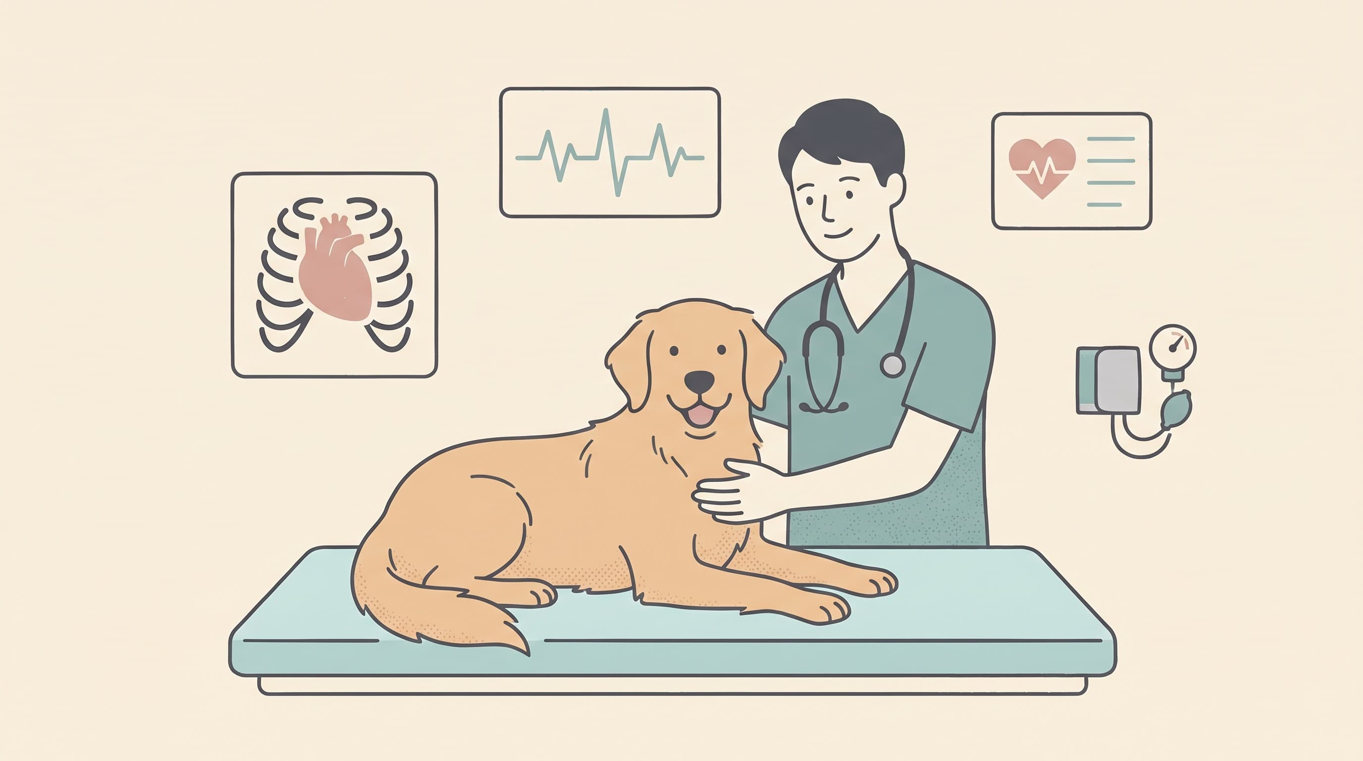
If your vet has heard a murmur, found an odd rhythm, or simply wants to check an older pet's heart, the next step is usually a test or two. Suddenly you are presented with a list: a chest x-ray, an echo, an ECG, a blood pressure check, a proBNP. It can feel like an alphabet of expense, and it is rarely explained well in the few minutes of a consult. This article is the plain-language version. For each test, here is what it actually looks at, what to expect on the day, and the honest answer to the question that really matters to you: will this one change the plan?
It helps to know how they fit together first, because it stops the list feeling random. No single heart test does everything. The x-ray shows the heart's size and the state of the lungs, the echo shows its structure and function, the ECG shows the electrical rhythm, the blood pressure shows the load the heart pumps against, and the blood tests show how hard the muscle is straining. Your vet is not ordering them to be thorough for its own sake. They are picking the few that answer the specific question your pet poses.
Chest x-ray: heart size and fluid in the lungs
The chest x-ray, or thoracic radiograph, is often the first imaging step, and it answers two big questions cheaply: is the heart enlarged, and is there fluid building up in or around the lungs. That second question is the one that turns a worry into an emergency, because fluid on the lungs, known as pulmonary oedema, is the hallmark of congestive heart failure.
To judge heart size objectively rather than by eye, vets often use a measurement called the vertebral heart score, which compares the heart against the length of the backbone, giving a number that can be tracked over time (Buchanan and Bücheler, 1995). It is a useful yardstick, though some perfectly normal breeds, certain Cavalier King Charles Spaniels and Boxers among them, naturally sit at the higher end, so the number is read in context rather than as a verdict.
On the day, an x-ray is quick and usually needs no sedation, though a wriggly or breathless patient may get a light sedative to keep the pictures sharp. Your pet lies on the table for a few seconds per view, normally two views, and that is it.
The honest limitation is this: an x-ray shows the heart's silhouette, the outline, but not the inside. It can tell you the heart is big, but not why, whether that is a leaky valve, a thick wall or a weak pump. For that, you need the echo. And in cats especially, the heart can look near enough normal in size on film while still being seriously diseased, which is why a normal chest x-ray is reassuring about fluid but cannot, on its own, give a cat's heart a clean bill of health.

Echocardiogram: the gold standard
If there is one test that earns the phrase "the gold standard", it is the echocardiogram, an ultrasound scan of the beating heart. Where the x-ray shows a shadow, the echo shows the working organ in real time: the thickness of each wall, the size of each chamber, how strongly the muscle squeezes, and, with the colour-Doppler setting, the direction and speed of blood flowing through the valves. It is the test that actually names the disease rather than just flagging that something is wrong. For the great majority of heart conditions in dogs and cats, the echo is what gives the definitive diagnosis (Côté et al., 2011).
It is also the test that most often changes the plan, and frequently in a way that directly affects your pet's lifespan. The clearest example is in dogs with the common leaky-valve disease, mitral valve disease. The landmark EPIC trial showed that starting the medication pimobendan in dogs whose hearts had already enlarged to a defined degree, but before any symptoms appeared, delayed the onset of heart failure by roughly fifteen months (Boswood et al., 2016). The catch is that the threshold for "enlarged enough to treat" is measured on the echo. Without the scan, you cannot know whether your dog has reached the point where early treatment buys that time. That is why a cardiologist will so often recommend an echo for a dog with a murmur: it is the difference between starting a proven treatment at the right moment and missing the window.
On the day, an echo is non-invasive and well tolerated. Your pet lies, usually on their side, often on a padded table with a cut-out so the probe can reach from underneath, and a patch of fur over the chest is clipped so the gel makes good contact. Most pets need no sedation at all, just gentle handling; a tense or fractious cat may be offered a mild calming medication. It takes anywhere from twenty minutes to the better part of an hour. There are no needles into the heart and no radiation, and your pet goes home the same day, none the wiser.
The one real friction is expertise. A heart scan is only as good as the person interpreting it, and a full diagnostic echo with accurate measurements is a genuine skill. Many first-opinion practices can perform a useful screening scan, but a referral to a cardiologist for a detailed study is often worth it when the result will drive a major decision, such as whether to start lifelong medication. We cover what a referral involves, and how to make the most of it, in working with a cardiologist.
ECG and Holter: reading the rhythm
An echo shows the heart's plumbing and pump. An electrocardiogram, the ECG or EKG, shows its electrics: the rhythm. It does not image the heart at all. Instead, small clips or pads on the skin pick up the electrical signals that coordinate each beat, drawing them as a trace. Its job is to answer a different question from the echo: is the heartbeat regular, too fast, too slow, or chaotic?
You reach for an ECG when the heart's rhythm, rather than its structure, is the concern, for instance an irregular or racing pulse, unexplained weakness, or fainting episodes. Some abnormal rhythms, called arrhythmias, are harmless quirks; others are dangerous and very treatable once identified, which is exactly why pinning them down matters. A resting ECG in the consulting room takes only a few minutes, needs no sedation, and is painless: your pet simply lies still while the trace is recorded.
The difficulty with rhythm problems is that the troublesome beats are often intermittent, and they have an unhelpful habit of behaving themselves for the five minutes your pet is wired up at the vet. This is where the Holter monitor comes in: a small, lightweight ECG recorder, worn in a little vest, that captures every heartbeat continuously for twenty-four hours or more while your pet goes about normal life at home (Côté et al., 2011). It is the right tool when symptoms come and go, because it catches the rare event the in-clinic snapshot misses. You keep a brief diary of any episodes, and the recording is analysed afterwards. In some breeds prone to inherited rhythm disease, the Boxer and the Dobermann especially, a Holter is also used to screen for early electrical changes before any structural disease is obvious.
Blood pressure: the load the heart pumps against, and why it matters most in cats
Measuring blood pressure in a pet is much like in a person: an inflatable cuff, usually on a leg or the tail, with the reading taken gently while your pet sits quietly. It is quick, painless and needs no sedation. What it tells you is the pressure the heart is working against, and that matters for the heart in two directions.
High blood pressure, hypertension, makes the heart's job harder and over time can thicken its walls and damage other organs, the eyes and kidneys in particular. It is common in older cats, where it usually rides along with another condition such as kidney disease or an overactive thyroid (Acierno et al., 2018). This is why a blood pressure check is so often folded into the workup for an older cat: catching and treating it can prevent a great deal of downstream harm, including sudden blindness, which is sometimes the first sign an owner ever notices.
The honest caveat is the white-coat effect, the same nervous spike that humans get at the doctor. A single high reading in a frightened animal can be misleading, so a careful clinic will take several readings in a calm, quiet room and let your pet settle first. One stressed number on its own is not a diagnosis of hypertension.
NT-proBNP and troponin: the strain blood tests
The newest tools in the box are blood tests, and they answer a question the imaging tests cannot: how hard is the heart muscle actually straining, right now? The main one is NT-proBNP, a substance the heart releases into the blood when its walls are stretched and stressed. A high level is a chemical signal that the heart is under load, well before you might see it on a scan.
Its great strength is as a triage and screening test, and it is especially valuable in cats. A common and genuinely difficult scenario is a cat brought in breathing fast and hard: is this a heart problem, or a lung or airway problem such as asthma? The two can look identical at the cage door but need opposite treatments, and a sick cat may be too fragile to scan safely until stabilised. Here a proBNP measured on the spot helps point the way, because a clearly high result makes heart failure much more likely and a low one makes it much less so (Fox et al., 2009; Côté et al., 2011). There is also an in-clinic SNAP-format version giving a fast positive-or-negative answer, useful as a quick filter, though the laboratory measurement is the more reliable number. A second marker, cardiac troponin I, rises when heart muscle cells are actually being damaged and can add supporting information in some cases.
Two honest limits keep these tests in proportion. They are a signal of strain, not a diagnosis: a high proBNP tells you the heart is stressed but not the cause, so it usually prompts an echo rather than replacing one. And results can be nudged by other things, kidney function among them, so they are read alongside the rest of the picture, never in isolation. Think of proBNP as a smoke alarm: excellent at telling you to look, not designed to tell you exactly what is burning.

What a "normal" or "abnormal" result really tells you
This is the part that causes the most quiet anxiety, so it deserves saying plainly. The most important thing to understand about every test above is what it does, and does not, rule out.
A normal result is reassuring, but it is specific to that one test. A normal chest x-ray means no obvious enlargement and, crucially, no fluid in the lungs today, which is genuinely good news in a breathless pet, but it does not mean the heart's internal structure is normal, because the x-ray never looked inside. A normal in-clinic ECG means the rhythm was regular during those few minutes, not that an intermittent arrhythmia can never happen, which is exactly why a Holter exists. A normal proBNP makes significant heart strain less likely but does not by itself exclude every cardiac problem. The only test that comes close to genuinely excluding structural heart disease is a thorough echo by an experienced operator, and even that is a snapshot of a disease that can progress.
The flip side is just as important, and often more comforting. An abnormal result is very often not a crisis. A heart that measures enlarged on an echo but is causing no symptoms may simply mean it is time to start a medication that buys months or years, as in the EPIC dogs (Boswood et al., 2016). A murmur that prompted all of this may turn out to sit alongside a structurally fine heart. An abnormal finding is information that sharpens the plan, not, in itself, a sentence. The whole purpose of testing is to replace a vague fear with a specific picture, and a specific picture is nearly always easier to live with, and to act on, than the worry that sent you in.
Whatever the results show, the most powerful tool for watching a heart over time is not a test at the vet at all, it is a number you can track at home for free. Counting your pet's breathing rate while they sleep is the earliest warning you have that fluid is starting to build, and it is the thread that ties all of this together between appointments. Our guide to the resting respiratory rate shows you exactly how to count it and what is normal, and you can log it with the breathing rate tracker. If you arrived here from a murmur, the what a murmur means article is the natural companion to this one, and if you are still getting your bearings on the heart itself, start with how your pet's heart works.
Keep track of how your pet is doing
The owners who cope best are the ones who notice changes early. A simple health log shows you what is working, and what is not, before the next vet visit.
Start tracking, freeYou're not doing this alone
Compare treatment journeys and talk to owners managing heart health. Free to join.
Join PetsLikeMine