
How to check your pet at home, and what to photograph
Alastair Greenway
BVM&S MRCVS
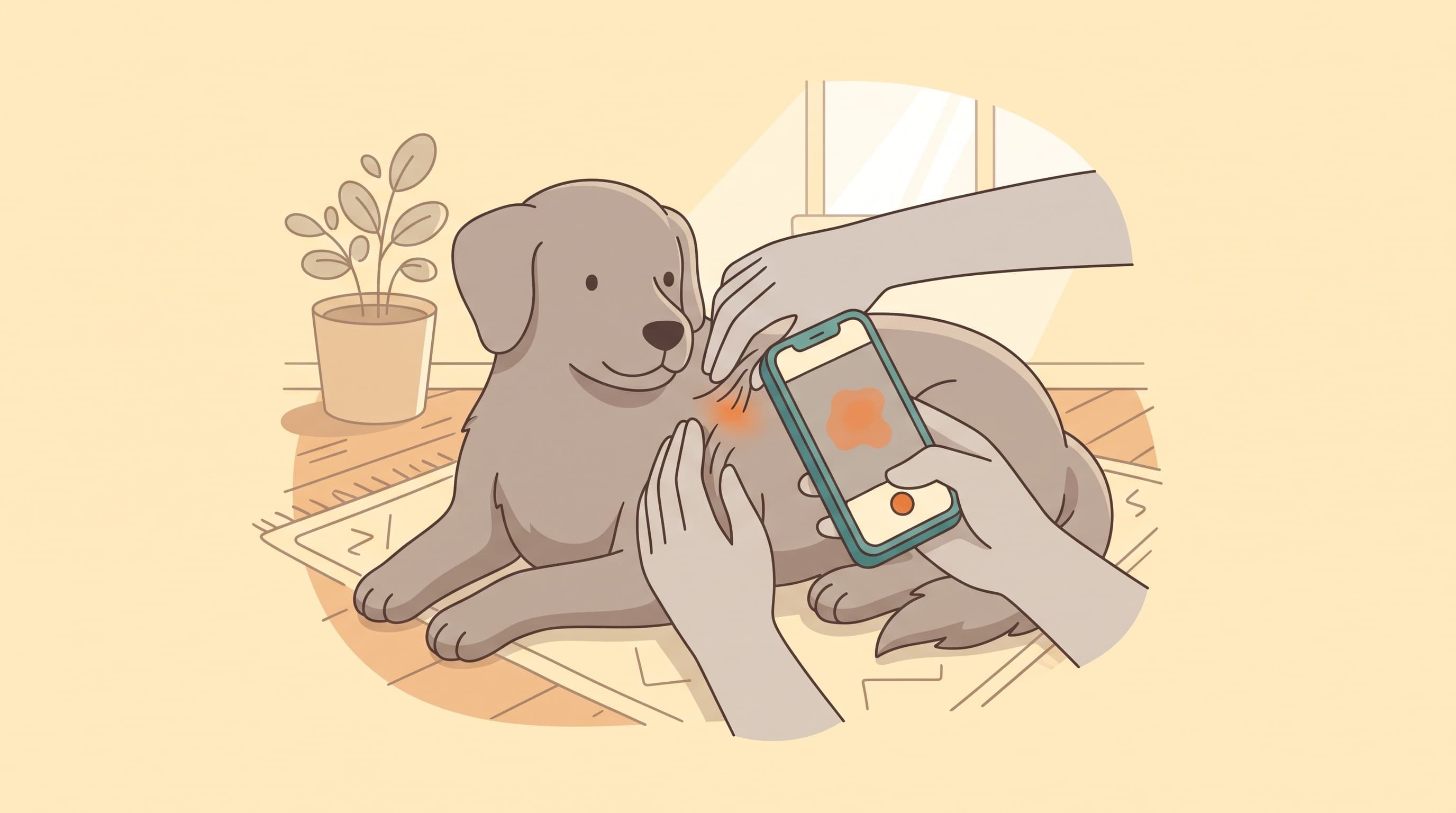
You see your pet every day. Your vet sees them for about ten minutes, on a day that may not be the day the skin looks its worst. That gap is the whole problem with skin disease, because itchy skin comes and goes, and a single look in the consulting room misses the pattern. So the most useful thing you can do is become a good observer: a quick check you can repeat, an itch score, and a couple of progress photos a vet can actually use. Owner-recorded itch is the validated way to capture what happens between visits and to judge whether treatment is working (Hill et al., 2007; Rybnicek et al., 2009). Do that, and your next appointment starts from real data instead of "she has been a bit itchy lately".
This guide is about the how: where to look, what to feel and smell for, how to score, and how to take a photo worth taking. What each finding actually means belongs to the work-up, so we hand that off to why is my pet so itchy and the section articles. Your job at home is to observe and record.
A simple head-to-tail route
Use the same route every time so nothing gets skipped. Start at the ears, then the face and muzzle, then the paws and between every toe, then the armpits (the axillae), the belly and groin, the rump and tail base, finishing with a hand through the coat and into any skin folds. There's a reason for that order. In dogs with atopic dermatitis the most commonly affected sites are the paws, face, ears, front-leg flexures, armpits and belly (Favrot et al., 2010), and flea-allergic dogs concentrate their misery over the rump and tail base while cats show it more around the head, neck and belly (Merck Veterinary Manual). A route that lingers in those places checks the high-yield ground first.
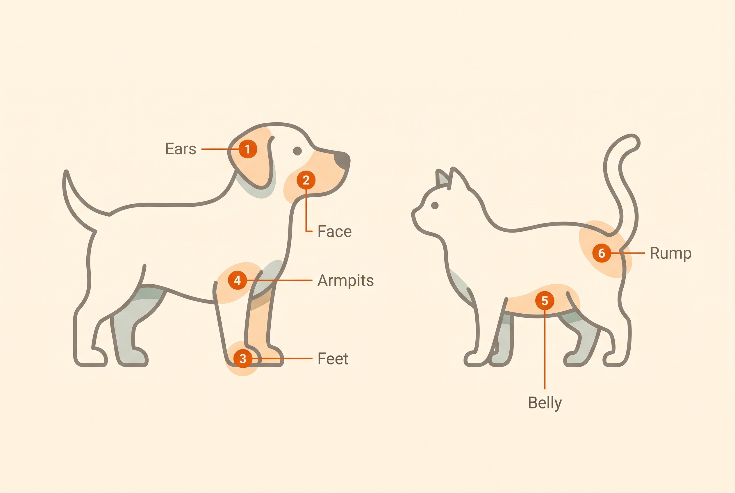
Do it when your pet is relaxed and in good light, during a normal cuddle so it does not feel like an examination. Lift the ear flaps, part the fur on the belly and inner thighs, and spread the toes to see the webbing. Most pets put up with this happily once it becomes routine, and a minute is all it takes. If your pet suddenly guards or snaps at one area, note it; pain is a finding too.
Look, feel and smell
The findings that matter are within reach of a careful owner, and you have three senses to use, not one. Owners tend to rely on their eyes alone and miss half of what is there. Look for redness (erythema), spots and pimples (papules and pustules), scabs and crusts, and the small circular rings of peeling skin left by a healed pustule (epidermal collarettes) (Favrot et al., 2010; Hillier et al., 2014). Look for thinning or bald patches, and for skin that has gone thickened, leathery and darker from months of rubbing ("elephant" skin). The distribution is worth noting in its own right, because the pattern is a clue a vet reads (Favrot et al., 2010).
Feel as you go. Healthy skin is supple and dry, so notice warmth, thickening, or a greasy, sticky feel that was not there before. Then smell. A yeasty, musty, almost corn-chip odour, often with greasy brown waxy debris matting the base of the hairs in warm folded areas, is the classic signature of Malassezia yeast overgrowth (Bond et al., 2020). Record these and show your vet; don't treat them yourself, because the infections are owned by skin and ear infections.
Here's the part most "check your pet's skin" pages skip. You cannot tell atopy from a food allergy from mange by looking. They overlap, they stack, and the same red itchy paw can have several causes at once. Even the formal clinical criteria are a pattern-recognition aid, good but not a diagnostic test, with a sensitivity around 85% and specificity around 79% at the usual cut-off of five of eight criteria (Favrot et al., 2010). So resist the urge to diagnose from the descriptors above; they exist to feed the work-up, not replace it.
The flea-dirt test, a real home diagnostic
There is one genuine test you can do at home, and it's worth learning, because flea allergy is the cheapest cause to rule out in any itchy pet. Comb the coat, concentrating over the rump and tail base in dogs and the head, neck and belly in cats, and tap the debris onto damp white paper or wet cotton wool. Flea dirt is digested blood, so the specks smear and dissolve to a rusty reddish-brown; ordinary grit stays grey or black (Merck Veterinary Manual). This matters because live fleas often go unseen even when present, so flea dirt is your proof of exposure (Dryden, 2009). A positive smear points to fleas. The "one bite is enough" story and the eradication plan belong to flea-allergy dermatitis and flea control.
Scoring the itch
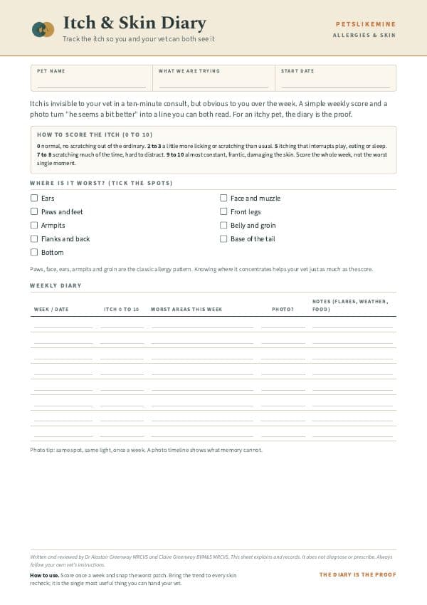
A 0 to 10 itch score is not a guess, it is a validated instrument. The pruritus Visual Analog Scale is the only owner-completed itch tool formally developed and validated in dogs, running a continuous line from a normal, healthy pet up to extremely severe itching all the time, anchored to real behaviours (Hill et al., 2007; Rybnicek et al., 2009). Two things make it trustworthy, and both are easy to copy.
First, score the behaviour, not just the obvious scratch. The validated scale counts licking, chewing, nibbling, rubbing the face, head-shaking and scooting as itch (Hill et al., 2007). Owners reliably under-call paw-licking and ear-rubbing, so add them in deliberately. Second, normal is not zero. In the validation cohort, plenty of dogs with no skin disease still scored above zero (90 of 305 healthy dogs), setting a normal range of roughly 0 to 1.9 (Rybnicek et al., 2009). A low background of scratching is normal, so the goal is to spot a rising trend, not to panic at a single scratch.
The actual scoring interface, the validated scale you slide and the trend it builds, lives in the Skin & Itch Tracker, where the whole home check is heading. Score the same way each time, log the site and date alongside the number, and a pattern surfaces: worse after walks, worse in late summer, always one ear. That is exactly the dataset a dermatology consult and a diet trial need. For why the score rises and falls, see the itch threshold.
Cats hide it, and they hide it differently
Cats are the reason a calm-looking pet can still be very itchy. They show itch mainly through licking and scratching, and much of the licking happens out of sight, so it is routinely under-reported (Colombo et al., 2022). The validated feline scale deliberately uses two separate lines, one for licking and one for scratching, taking the higher as the score, because in cats those behaviours measure genuinely different things (Colombo et al., 2022). So don't wait to witness the grooming, because you often will not.
Read the coat instead. The tell of overgrooming in cats is hair broken off short that feels stubbly against your hand, leaving thinned or bald areas with skin that often looks normal underneath, so-called barbered or self-induced alopecia (Colombo et al., 2022; Halliwell et al., 2021). Run a hand against the lie of the coat on the belly, flanks and inner thighs and feel for that stubble; if you find it, treat it as overgrooming until proven otherwise. Telling true hair loss from a barbered coat, and itch from a behavioural cause, is owned by feline atopic syndrome.
What and how to photograph
A progress photo is only worth taking if it can be compared, and that comparison falls apart the moment the lighting, distance and angle drift. Owner-collected images are a genuinely useful monitoring tool once they are standardised (Strzok et al., 2022). A few rules get you there:
- Same spot, same light. Daylight near a window beats yellow indoor bulbs, and both beat flash, which flattens texture and shifts the colour. Photograph the same patch in the same light each time, flash off.
- Get close and square-on. Hold the camera roughly parallel to the skin rather than at an angle, fill the frame, and have a second person part the fur. Steady your elbow so the shot is in focus.
- Include scale and a date. Put something familiar in shot for size, a coin, a ruler, or a finger beside the lesion, mirroring how a clinic standardises measurements. Note the date and where on the body it is.
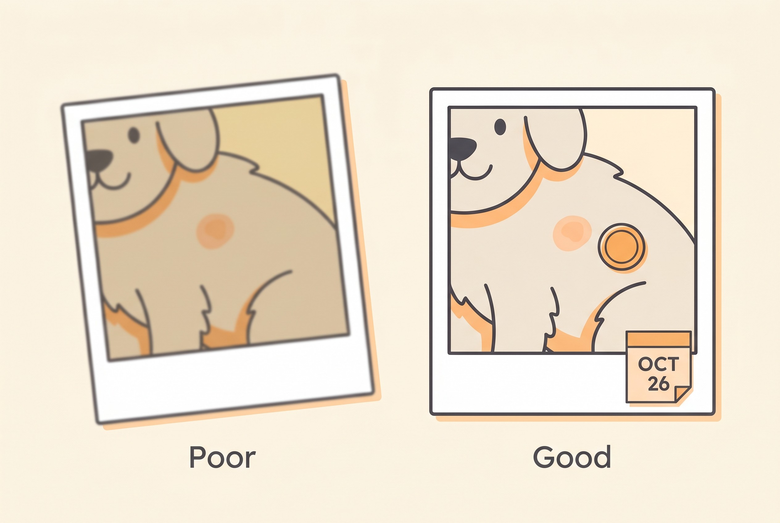
Take two kinds of picture each time. A close-up of the worst patch tracks that lesion; a wider shot captures the whole distribution, which armpits, which feet, the belly, the tail base, because the pattern itself is diagnostic information (Favrot et al., 2010). The same habit works for any new lump, where a record of site, size and date lets a vet see whether it is changing; that triage is owned by lumps, bumps and spots.
Record it like data, not a diary
For every finding that catches your attention, capture five things: where it is (the site), what it looks like, how big it is, since when, and the itch score for the day. "Itchy again this week" tells a vet nothing; "left front paw, red between the toes, licking it, score 6, started four days ago" tells them a great deal, because that is the precise shape an elimination-diet trial and a dermatology referral both run on. Consistent home scoring is, in fact, how you read a food trial: track the score across eight strict weeks and the number is your readout, which is why the trial design and a companion to run it live in the elimination diet done properly and the Elimination-Diet Companion.
When to stop logging and pick up the phone
A home check is screening, not diagnosis, and most of what you find is something to log and watch. A few things are not. A hot, fast-growing, painful patch; sudden facial swelling or hives; a cat breathing oddly; or a suddenly very painful ear with a head tilt are not for the tracker, they are reasons to act now. Telling the genuinely urgent from the merely miserable is owned by skin emergencies, with a fast triage at the itchy-pet checker.
For everything else, you have already done the hard part. Do the first check tonight, give the itch a score, take the wide-then-close photo pair with a coin and the date in shot, and log it in the Skin & Itch Tracker. That single record is more than most consultations ever start with, and from your second check on you are no longer guessing at a trend, you are watching one.
References
- Hill PB, Lau P, Rybnicek J. Development of an owner-assessed scale to measure the severity of pruritus in dogs. Veterinary Dermatology. 2007;18(5):301-308.
- Rybnicek J, Lau-Gillard PJ, Harvey R, Hill PB. Further validation of a pruritus severity scale for use in dogs. Veterinary Dermatology. 2009;20(2):115-122.
- Colombo S, Sartori R, Schievano C, Borio S. Development and validation of an owner-assessed Visual Analog Scale for feline pruritus severity scoring (VAScat). Veterinary Dermatology. 2022;33(5):407-413.
- Favrot C, Steffan J, Seewald W, Picco F. A prospective study on the clinical features of chronic canine atopic dermatitis and its diagnosis. Veterinary Dermatology. 2010;21(1):23-31.
- Bond R, Morris DO, Guillot J, Bensignor EJ, Robson D, Mason KV, Kano R, Hill PB. Biology, diagnosis and treatment of Malassezia dermatitis in dogs and cats: Clinical Consensus Guidelines of the World Association for Veterinary Dermatology. Veterinary Dermatology. 2020;31(1):28-74.
- Loeffler A, Cain CL, Ferrer L, Nishifuji K, Varjonen K, Clark SM, Bergvall K; ISCAID. Antimicrobial use guidelines for canine superficial bacterial folliculitis (pyoderma) by the International Society for Companion Animal Infectious Diseases (ISCAID). Veterinary Dermatology. 2025. (Update to Hillier et al. 2014; doi to confirm.)
- Dryden MW. Flea and tick control in the 21st century: challenges and opportunities. Veterinary Dermatology. 2009;20(5-6):435-440.
- Merck (MSD) Veterinary Manual. Flea Allergy Dermatitis in Dogs and Cats. Integumentary System.
- Strzok E, Siettou C, Mueller RS, et al. Validation of the 0 to 10 verbal numeric scale for assessment of pruritus severity in dogs. Veterinary Dermatology. 2022;33(3):209-216.
- Halliwell R, Pucheu-Haston CM, Olivry T, Prost C, Jackson H, Banovic F, Nuttall T, Santoro D, Bizikova P, Mueller RS. Feline allergic diseases: introduction and proposed nomenclature. Veterinary Dermatology. 2021;32(1):8-e2.
Free downloads
Companion worksheets to put what you've read into practice. Free PDFs, print at home.
Keep track of how your pet is doing
The owners who cope best are the ones who notice changes early. A simple health log shows you what is working, and what is not, before the next vet visit.
Start tracking, freeYou're not doing this alone
Compare treatment journeys and talk to owners managing allergies & skin. Free to join.
Join PetsLikeMine