Lumps, bumps and spots: which need the vet now, and which can wait
Dr. Alastair Greenway
MRCVS
You are stroking your pet, half-watching the television, and your fingers stop on something that was not there before. A pea-sized lump under the skin. The mind goes to the worst place, and then to the internet, where you will read that soft and movable means fatty and harmless, while firm and fixed means trouble. That advice is comforting, widely repeated, and wrong in the way that matters most. This article is about what a new lump might be, when to worry, and the single calm, cheap step that answers the real question. It is not a diagnosis tool: lumps cannot be diagnosed at a distance, so its job is to get you to the right appointment with the right information.
Why you cannot tell by look or feel
Here is the uncomfortable truth, and it comes from the people best placed to know it. In the WSAVA's practical guidance on lumps and bumps, the veterinary oncologist Dr Sue Ettinger puts it plainly: even the most experienced vet or oncologist cannot look at or feel a mass and know whether it is malignant, because cancer is a cellular diagnosis (Ettinger, WSAVA 2017). Size, softness, colour, whether it slides freely under the skin, none of it reliably sorts the harmless from the dangerous, because benign and malignant masses overlap in exactly those features. A lump that feels for all the world like a harmless fatty lump can be a soft-tissue sarcoma or a mast cell tumour sitting deep under the skin. So when a website tells you a soft, movable lump is "probably nothing", it is making a guess the best oncologists in the world will not make with their hands. The useful question, then, is not what a lump looks like but how to find out cheaply.
The cheap answer: a fine-needle aspirate
The answer is a fine-needle aspirate, usually shortened to FNA. A thin needle goes into the lump, a few cells are drawn off, smeared on a slide and looked at under the microscope, often within minutes and often in the consulting room without sedation. Its advantages are what a worried owner wants to hear: minimally invasive, low risk, low cost, with results far faster than a surgical biopsy (Ettinger, WSAVA 2017).
It is worth being honest about how good it is. In the largest correlation study relevant to UK practice, covering 242 dogs and 50 cats, the cells from an aspirate agreed with the final tissue diagnosis in 90.9% of masses, correctly flagging a tumour with 89.3% sensitivity and 97.9% specificity (Ghisleni et al., 2006). A clear result is usually trustworthy. It is not perfect, though: some masses, certain sarcomas in particular, do not shed cells well onto a needle, so a "no tumour cells seen" result does not rule cancer out the way a positive result rules it in (Ghisleni et al., 2006). When the cells are inconclusive, the vet moves on to a biopsy. The aspirate sorts most lumps and tells the vet whether that bigger step is needed.
The "watch and wait" trap
The most common advice an owner gives themselves is "I'll keep an eye on it". The trouble is that a mass which turns out to be cancer does not get easier to treat while you watch. As a tumour grows, what could have been lifted out in a small, often curative operation may come to need a much bigger surgery, sometimes with radiation or chemotherapy afterwards (Ettinger, WSAVA 2017). Catching it small is what makes a clean removal likely.
So there is a simple rule, from the campaign Ettinger named "See Something, Do Something. Why Wait? Aspirate": if a lump is the size of a pea, around one centimetre, and has been there a month, get it sampled (Ettinger, WSAVA 2017). That is the single most useful number here. It does not mean rushing to the emergency clinic, just booking a routine appointment for a quick sample.

For reassuring context, most skin lumps in dogs are not cancer. In the Danish national cancer registry, 66% of canine skin tumours were benign against 21% malignant, and a northern Portuguese laboratory series found 62.9% benign against 37.1% malignant (Brønden et al., 2010; Martins et al., 2022). The aspirate is not because a lump is probably sinister, but because it is the only cheap way to be sure.
The common, usually benign ones
A handful of lumps turn up again and again, and it helps to know them, as long as you hold on to the rule that even the friendly-sounding ones earn one check.
Fatty lumps (lipomas) are common, soft, benign tumours of fat, especially in older and heavier dogs (Martins et al., 2022). They are also the exact reason "it's just a fatty lump" is a risky assumption, because a soft subcutaneous mast cell tumour or a soft-tissue sarcoma can feel identical on the fingertips, so the classic fatty lump still earns one aspirate to confirm it really is fat (Ettinger, WSAVA 2017).
Warts (viral papillomas) are small, often cauliflower-like growths caused by papillomavirus, classically around the mouth and on the skin of young dogs whose immune systems are still maturing. Most are benign and regress on their own within about three months as immunity develops, and they are highly species-specific, so they pass between dogs but not to people (Medeiros-Fonseca et al., 2023).
Sebaceous and follicular cysts are blocked oil glands or hair follicles that fill with sebum or keratin. They are usually benign and slow, but a burst cyst spills its contents and sets off a brisk, sometimes painful inflammatory reaction (VCA Animal Hospitals).
Histiocytomas are the young dog's "button tumour": a benign growth of Langerhans cells seen mostly in dogs under two, typically a single fast-growing, hairless, raised button on the head, ears or limbs that regresses on its own within a couple of months as the dog's own immune cells clear it (Diehl and Hansmann, 2024; VCA Animal Hospitals). This is one of the few lumps where a vet may advise watching, but only after an aspirate has confirmed what it is, because a fast-growing lump in a young dog could also be something else entirely.
The ones that earn the quick sample
Mast cell tumours are the reason this whole article exists. They are the most common malignant skin tumour in dogs, somewhere around one in six skin tumours in many series, and they have earned the nickname "the great pretender" because their appearance is so variable: a wart-like nodule, a soft lump that feels just like a lipoma, or an angry ulcerated mass, so appearance alone cannot establish the diagnosis (de Nardi et al., 2022). They can even swell and redden when handled, because squeezing them releases histamine. The good news pairs with the warning: they shed cells easily and have a distinctive look down the microscope, so a fine-needle aspirate is usually enough to diagnose one (Willmann et al., 2021). The grade, and how it will behave, still needs a surgical biopsy, but the flag goes up at the cheap first step.
Soft-tissue sarcomas are the other common malignant group, and, like deep mast cell tumours, they can pose as a soft, slow "fatty lump". They are one more reason never to dismiss a deep soft lump on feel alone, though their cells often do not characterise the mass fully, so a biopsy is usually needed (Ettinger, WSAVA 2017; Martins et al., 2022).
Cats are different, and the rules change
A great deal of lump advice is dog-shaped, and cats need their own. When a cat does grow a skin tumour, it is more likely than a dog's to be malignant: in a UK study of over 9,000 feline skin tumours, 52.7% of the neoplastic masses were malignant, with fibrosarcoma and squamous cell carcinoma high on the list alongside benign basal cell tumours (Ho et al., 2018). So the reassuring "most lumps are benign" line is a dog line. In cats, the threshold to have a new lump checked should simply be lower.
One cat-specific lump is worth knowing by name, calmly. An injection-site sarcoma is a firm lump that can develop where a cat has had an injection, a vaccine or otherwise, and persists or grows. The trigger to act is the widely used "3-2-1 rule": get a biopsy if a post-injection lump is still there at three months, becomes bigger than two centimetres, or is still growing one month after the injection (Hartmann et al., ABCD guideline; AAHA/AAFP). This is not a reason to fear vaccinating, because the diseases vaccines prevent are far more common and dangerous, and the sarcoma risk is well below one in 10,000 doses (AAHA/AAFP). It is a reason to feel injection sites afterwards and flag a persistent lump early, because early, wide surgery gives the best outcome (Hartmann et al., ABCD guideline).
When it is "book soon", "see today", or genuinely fine
For most lumps, the pea-and-a-month rule is your default: book a routine appointment for an aspirate (Ettinger, WSAVA 2017). Some features push it up to "book promptly", though not necessarily an emergency: a lump growing quickly, changing in size, shape or colour, ulcerated or bleeding, fixed to deeper tissue rather than sliding freely, or a sore that will not heal (Ettinger, WSAVA 2017). Any new lump on a cat belongs in this prompt category too (Ho et al., 2018).
A faster lane exists too. A lump that is suddenly hot, painful and swelling fast, or one that comes with a pet who is off their food, lethargic or unwell, may be an abscess or a deep infection rather than a tumour, and that is a same-day call. Sudden facial swelling, hives or any difficulty breathing is more urgent still. Acute swelling and skin emergencies are covered properly in is this a skin emergency, so this article points there rather than treading the same ground. In the same spirit, if what you found is really a pustule or a weeping, infected lesion, that is the territory of skin and ear infections, and a raw, suddenly-appeared moist patch is usually a hot spot, covered in hot spots in pets.
What to record while you wait for the appointment
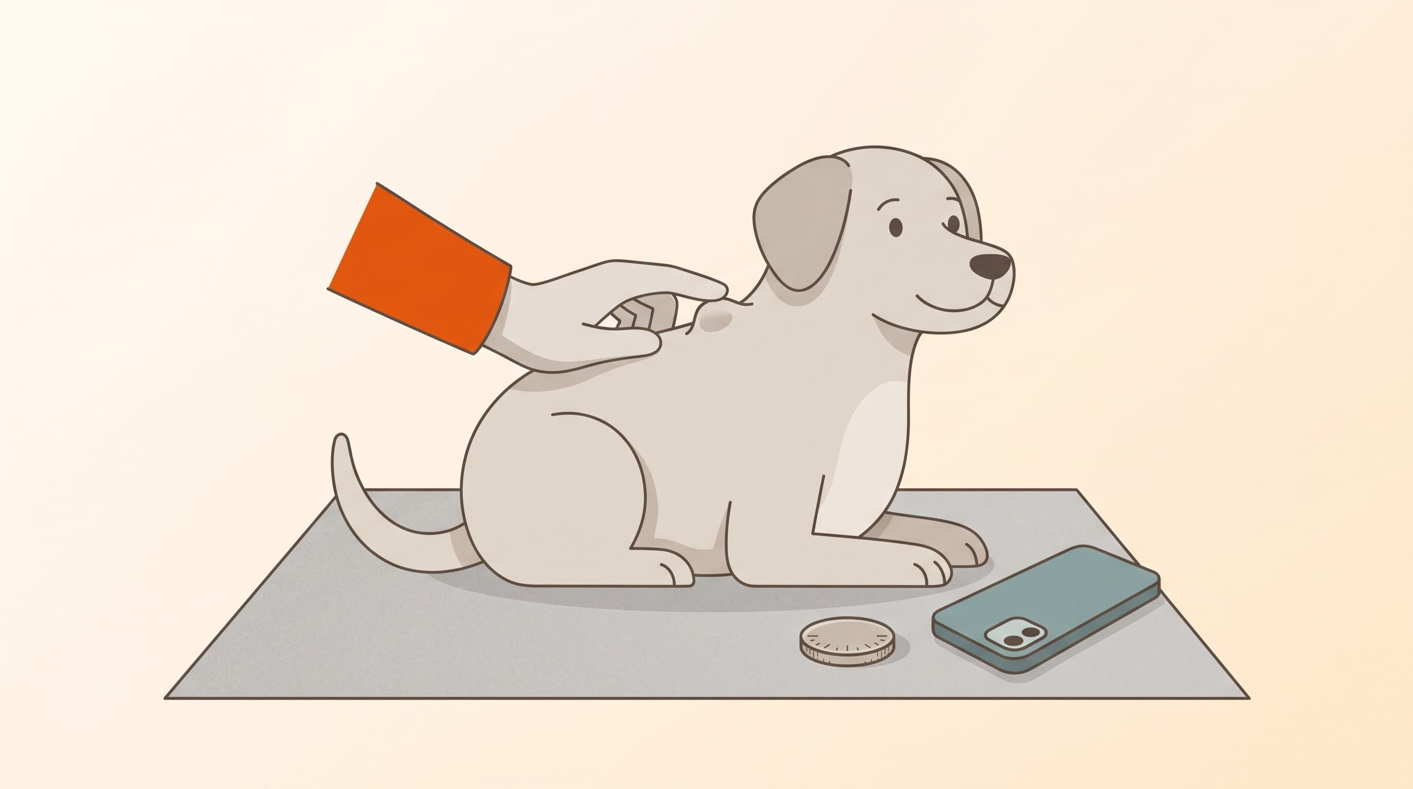
The genuinely useful thing you can do between finding a lump and the visit is to measure and document it, because that turns "I think it's grown" into evidence the vet can act on: a measurement across the widest point, a coin or ruler for scale, the exact location so the same lump is tracked, and a dated photograph (Ettinger, WSAVA 2017).
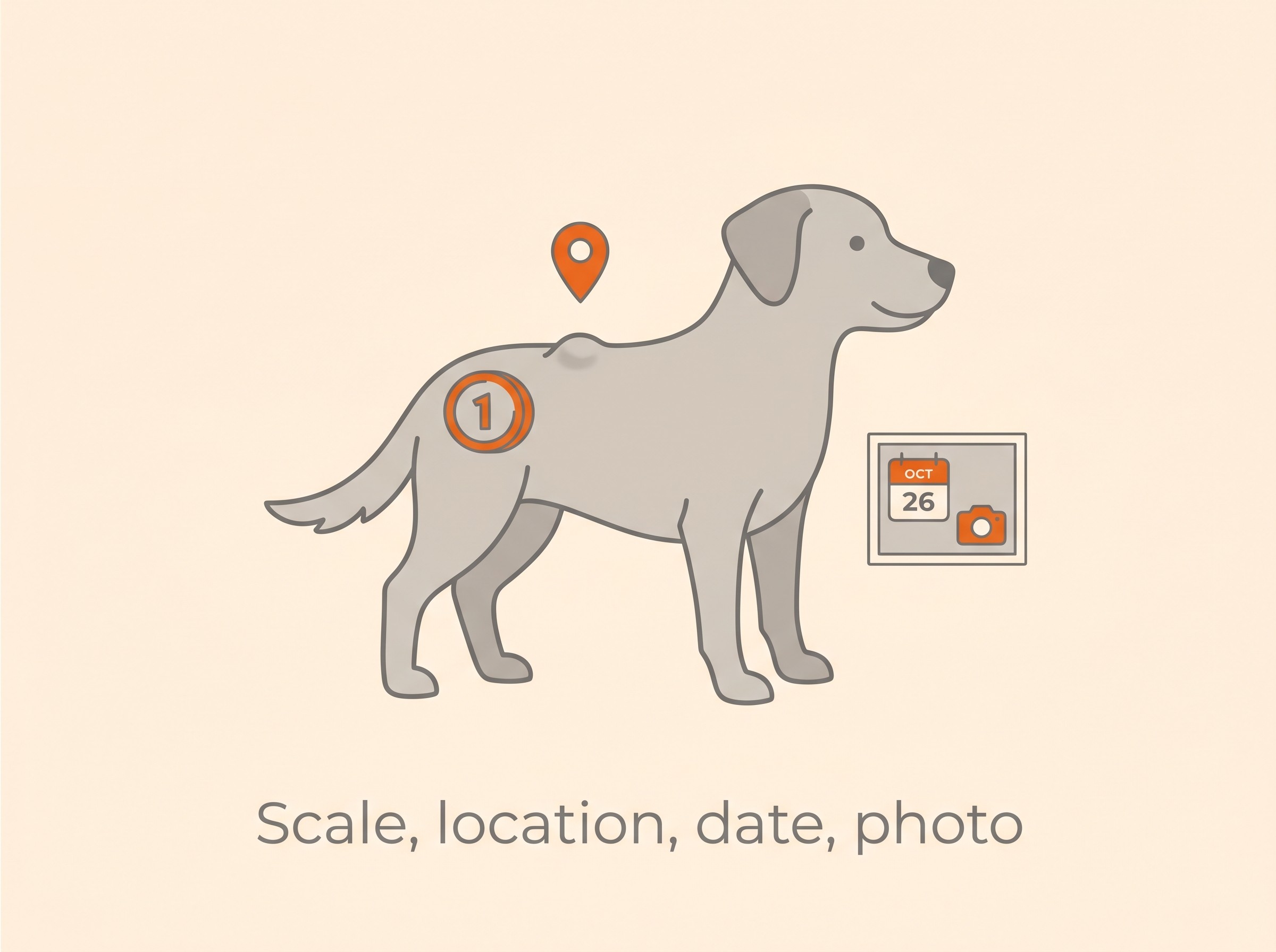
This is exactly what the Skin & Itch Tracker is built to hold: a dated record that gives your vet a growth curve and tells apart a stable old lump, unchanged for two years, from a creeping new one that has doubled in a month. If you found this on a routine going-over of your pet, how to check your pet's skin at home sets that habit out properly. The quickest way to stop worrying about the lump you found today is to let your vet draw off a few cells and tell you what it is.
Keep track of how your pet is doing
The owners who cope best are the ones who notice changes early. A simple health log shows you what is working, and what is not, before the next vet visit.
Start tracking, freeYou're not doing this alone
Compare treatment journeys and talk to owners managing allergies & skin. Free to join.
Join PetsLikeMine