The Diagnostic Workup: Bloods, MRI, CSF and What It Costs
Dr. Alastair Greenway
MRCVS
Your dog has had a seizure, or a few, and now there's a list of tests on the table. Bloods, maybe blood pressure, possibly an MRI under anaesthetic with a price tag that makes your stomach drop. You want to do right by your dog, but you also want to know what's genuinely necessary, what each test actually tells you, and whether you can stop at a sensible point without feeling you've cut a corner.
That's what this article is for. I'll walk you through the workup the way a vet builds it, one step at a time, with what each test rules in or out, the honest truth about the anaesthetic, and some UK cost ranges so the numbers aren't a mystery. Two boundaries. I won't re-argue why a pile of normal results can add up to a diagnosis, because that logic lives in why epilepsy is a diagnosis of exclusion. And I won't relitigate the causes of seizures, because reactive, structural and idiopathic seizures explained owns that ground. This piece is about the tests, what they cost, and how far to go.
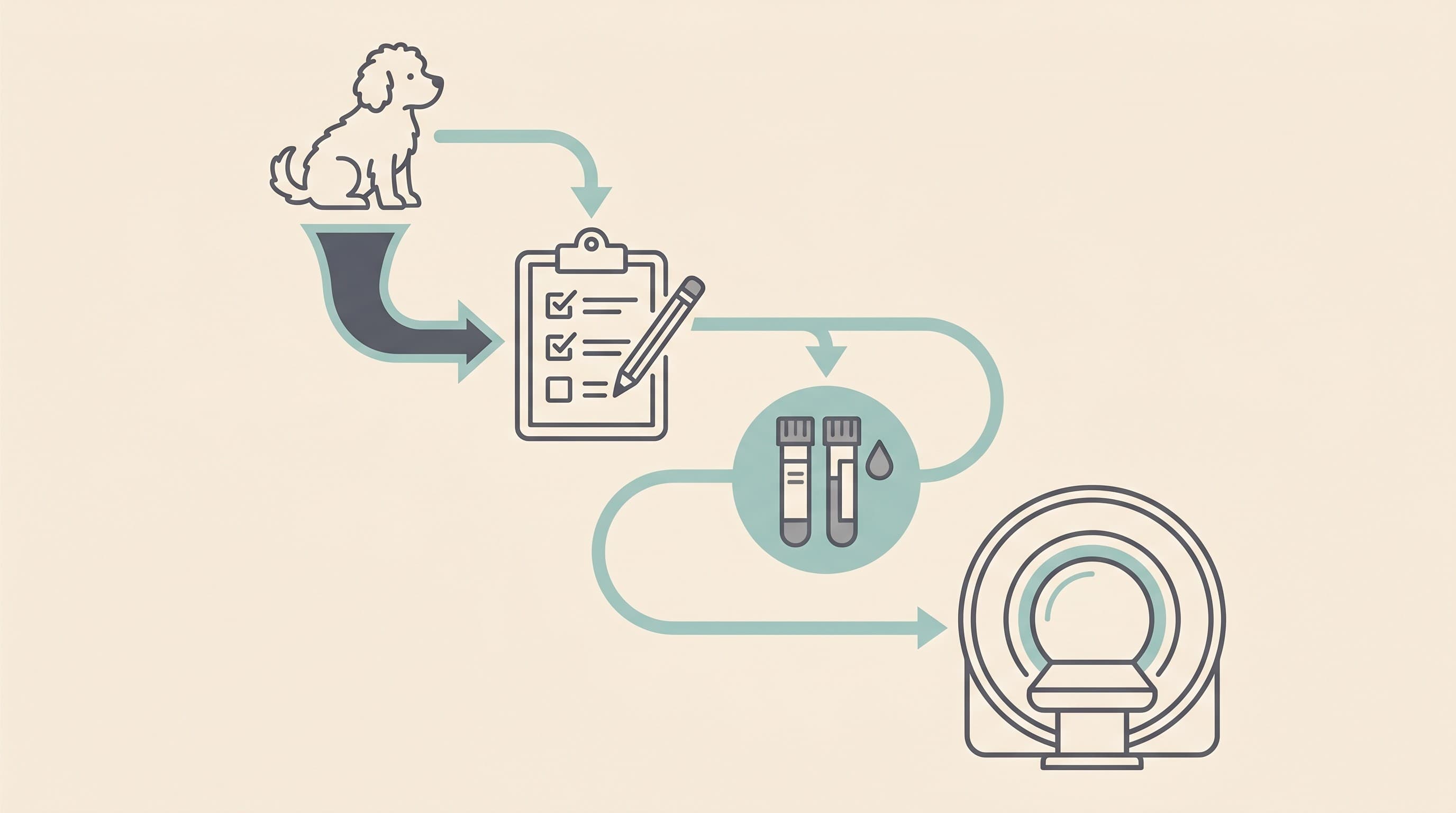
It starts before any test: history and examination
Here's the part people skip past, hunting for the scan. The single most useful investigation needs no machine at all. It's a good description of what happened, ideally a video, and a record of how often it's happening. The International Veterinary Epilepsy Task Force, the consensus group whose framework most vets follow, is explicit that the workup begins with a thorough history and a full physical and neurological examination, and that those two things drive every decision about which tests come next (De Risio et al., 2015).
This is where your homework genuinely changes your dog's care. Seizures almost never happen helpfully in the consulting room, so the vet is relying on you to be their eyes. Our seizure diary is built for exactly this: time the event, log the recovery, capture the pattern. That record isn't just admin. It feeds straight into how far down the testing road you'll need to travel, because a young dog with a textbook history and a normal exam is a very different proposition from an older dog whose exam isn't quite right.
The neurological exam matters because of what it predicts. A dog that's abnormal between seizures, in what vets call the interictal period, is far more likely to have something visible inside the brain than a dog that's normal between events. Hold that thought, because it becomes the hinge the whole "do we scan?" decision turns on.
The first tests: bloods, urine and what they're looking for
The starting database is bloods plus urine. The IVETF lists a complete blood count, a serum biochemistry profile and a urinalysis as the minimum (De Risio et al., 2015). The biochemistry panel includes sodium, potassium, chloride, calcium, phosphate, alanine aminotransferase (ALT), alkaline phosphatase (ALP), total bilirubin, urea, creatinine, total protein, albumin, glucose, cholesterol, triglycerides, and fasting bile acids and/or ammonia, while the urinalysis covers specific gravity, protein, glucose, pH and sediment cytology (De Risio et al., 2015). You don't need to memorise that. The point is that it's a wide net.
What's it catching? The things that make a perfectly healthy brain misfire: low blood sugar, electrolyte disturbances, kidney disease and, particularly, liver problems (De Risio et al., 2015). A seizure caused by one of these isn't epilepsy at all, it's a reactive seizure, and that distinction belongs to reactive, structural and idiopathic seizures explained. The practical upshot is that these cheap, low-risk tests can sometimes find a treatable cause and spare your dog the bigger workup entirely.
The liver gets special attention through the bile acids and ammonia. These check for liver dysfunction and for a portosystemic shunt, a blood vessel that bypasses the liver, both of which can poison the brain just enough to trigger seizures. It's why a young dog that starts seizing may be screened for a liver shunt before anyone mentions an MRI, with paired fasting and post-prandial bile acids forming part of the next, higher-confidence tier (De Risio et al., 2015). Blood pressure is part of the screen too, especially in older animals and cats, since hypertension is a recognised contributor and more relevant on the feline side (De Risio et al., 2015; Raimondi et al., 2017).
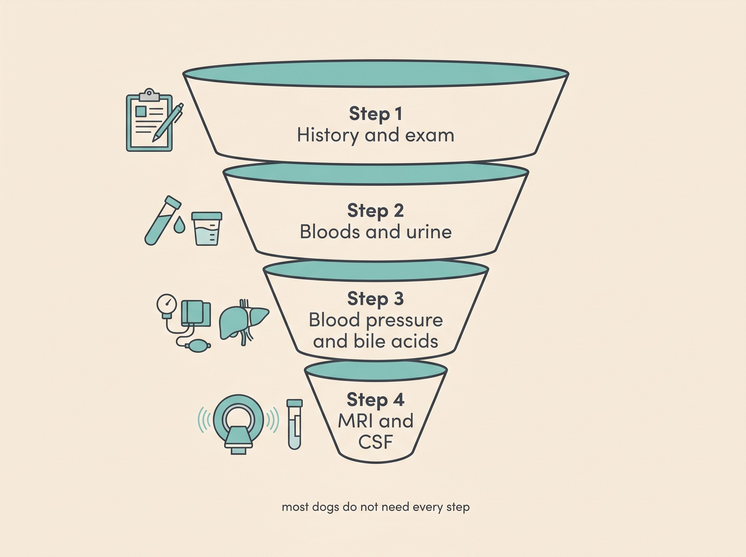
When MRI and a spinal tap genuinely earn their place
An MRI of the brain, usually paired with a sample of the fluid around it, is the advanced step. This is the one that looks inside the brain for a structural cause: a tumour, inflammation, a malformation, a stroke, or in cats a specific change called hippocampal necrosis. MRI is the imaging modality of choice for brain tissue (De Risio et al., 2015; Rusbridge et al., 2015).
The honest question is when it's worth it, because it's the expensive, anaesthetic-requiring step. The IVETF recommends advanced imaging in dogs with onset before six months or after six years of age, with interictal neurological abnormalities pointing to a problem inside the skull, with status epilepticus or cluster seizures at onset, or with a previous presumptive diagnosis that has since become drug-resistant despite a single drug pushed to its highest tolerable dose (De Risio et al., 2015). In plainer terms: very young or older at onset, an abnormal exam between seizures, a dramatic or clustering first presentation, or a dog not responding to a properly dosed drug. Those are the dogs in whom finding something is likely enough to justify the scan.
And here's the figure that stops owners assuming a normal exam means a normal brain. One study cited by the IVETF found brain MRI abnormalities in 22% (14/63) of epileptic dogs with a normal neurological examination, and in 90% (47/52) of those with an abnormal one (De Risio et al., 2015). So an abnormal exam makes a structural lesion very likely, and a normal exam makes it much less likely without quite ruling it out. Larger, more recent data refine that reassuringly: in 412 dogs with seizures and a normal interictal exam, only 16, just 3.9%, had a structural lesion (Phillipps & Goncalves, 2025). The catch is age, with the risk climbing sharply to around 23.1% in dogs over eight years (Phillipps & Goncalves, 2025). That's the evidence behind individualised advice: a young adult with a normal exam may reasonably start treatment without rushing to a scan, while an older-onset dog should be imaged.
Why the scan comes before the tap, and why the protocol matters
If your dog does go for an MRI, the spinal fluid sample, the CSF tap, is taken afterwards, and that order is a safety decision, not a scheduling one. Collecting CSF is dangerous when pressure inside the skull might be raised, because the tap itself can precipitate fatal brain herniation, where the brainstem is forced through the base of the skull. The MRI is done first precisely to check for a mass or signs of raised pressure before any fluid is drawn (De Risio et al., 2015). So the testing is deliberately sequenced for safety, never done blindly. One nuance if you're later told the CSF was "slightly abnormal": a recent seizure can itself cause a transient rise in white cells in the fluid, so results are read in context, not in isolation (Rusbridge et al., 2015).
The quality of the scan, and who reads it, matter more than most owners realise. The IVETF recommends an epilepsy-specific protocol of thin slices angled to the hippocampus in several planes, a particular set of image types, and a sequence sensitive to bleeding (Rusbridge et al., 2015). High-field scanners (1.5 Tesla or above) are preferred, yet a lot of veterinary MRI is still done on low-field machines that can miss small lesions (Rusbridge et al., 2015). Expertise counts too: in one analysis, 61% of epilepsy-causing lesions were missed when "standard" scans were reported by non-experts (Rusbridge et al., 2015). That's the real argument for having this done at a centre with a neurologist and a proper scanner, and part of why your vet may refer you. What that referral involves, and what to take with you, is covered in when your vet refers you to a neurologist.
The anaesthetic: small but real, and worth being honest about
An MRI and a CSF tap both need your pet fully anaesthetised and completely still, and no owner takes that lightly. So here are the actual numbers. The UK's large CEPSAF study put the overall anaesthetic-related death risk at roughly 0.17% in dogs and 0.24% in cats, falling to around 0.05% in healthy dogs and 0.11% in healthy cats, and rising in sick animals (Brodbelt et al., 2008). In a healthy patient that's genuinely small, on the order of one in two thousand, though never zero. A seizure patient is monitored especially carefully, because anaesthetic drugs and the recovery period interact with seizure risk. The way to weigh it is simple: a small, real risk, set against a scan that could change everything you do next. If the scan won't change the plan, that risk is harder to justify. If it will, it's usually worth it.
What it costs in the UK
A word before the numbers: every figure here is a practical estimate that varies widely by region, practice and date, and none of it is peer-reviewed. Treat it as a ballpark, not a quote.
The first-line bloods and urine are the affordable end, often tens to low hundreds of pounds. The MRI is the big cost. Independent UK guides put a dog brain MRI broadly in the region of £1,500 to £4,500 or more depending on the area, the scanner and whether contrast and an overnight stay are involved, with one 2025 estimate landing around £3,800 on average (NimbleFins, 2025). To make that concrete, Langford Vets (prices valid May 2026) lists a neurology consultation at around £340 and a full seizure investigation, MRI and CSF with the consult included, in the region of roughly £3,675 to £4,500, described as indicative and subject to change (Langford Vets, 2026). Ring your own practice for a real estimate. The bigger picture, lifetime medication, monitoring and how insurance handles a chronic condition, is its own subject and lives in the cost of epilepsy and how insurance works.
How far do we go? A framework, not a verdict
This is the part I most want you to take away, because it's where owners carry needless guilt. There is no single right amount to spend or test. The right level of investigation is the one that answers the question that would actually change what you do.
Reaching a confident diagnosis from the first tier alone, the right age, a normal exam and normal bloods, can be entirely enough to start treatment without an immediate MRI (De Risio et al., 2015). Choosing not to scan a textbook young-adult dog is a legitimate, common decision, not neglect. Equally, an atypical age at onset, an abnormal exam, a cluster or status presentation, or a poor response to a well-dosed drug are real reasons to image (De Risio et al., 2015; Phillipps & Goncalves, 2025). Both can be good medicine. What ties them together is one simple test: will this scan change the plan? If yes, it's worth considering. If no, you can stop with a clear conscience.
The same pattern in cats, only sooner
Cats follow the same logic with the dial turned up. In a study of 188 cats with seizures and a normal neurological exam, around 12% had MRI abnormalities overall, rising to 23.1% in cats over six years of age (Raimondi et al., 2017). Combined with the fact that structural and toxic causes are relatively more common in cats than in dogs, this is why vets recommend investigating a seizing cat sooner rather than waiting and seeing. The full feline cause list sits in what causes seizures in cats.
Whatever path you and your vet choose, the thread running through all of it is information: the better the history and diary you bring, the smarter every decision downstream becomes. So if you do one thing today, keep timing and filming the seizures and logging them in the seizure diary. The tests exist to answer specific questions, and a confident answer, even one that means "we can stop here", is the whole goal. When you're ready to think about what a referral involves, seeing a neurologist is the next step.
References
- De Risio, L., Bhatti, S., Muñana, K., Penderis, J., Stein, V., Tipold, A., Berendt, M., Farquhar, R., Fischer, A., Long, S., Mandigers, P. J. J., Matiasek, K., Packer, R. M. A., Pakozdy, A., Patterson, N., Platt, S., Podell, M., Potschka, H., Batlle, M. P., Rusbridge, C., & Volk, H. A. (2015). International Veterinary Epilepsy Task Force consensus proposal: diagnostic approach to epilepsy in dogs. BMC Veterinary Research, 11, 148.
- Rusbridge, C., Long, S., Jovanovik, J., Milne, M., Berendt, M., Bhatti, S. F. M., De Risio, L., Farquhar, R. G., Fischer, A., Matiasek, K., Muñana, K., Patterson, E. E., Pakozdy, A., Penderis, J., Platt, S., Podell, M., Potschka, H., Stein, V. M., Tipold, A., & Volk, H. A. (2015). International Veterinary Epilepsy Task Force recommendations for a veterinary epilepsy-specific MRI protocol. BMC Veterinary Research, 11, 194.
- Brodbelt, D. C., Blissitt, K. J., Hammond, R. A., Neath, P. J., Young, L. E., Pfeiffer, D. U., & Wood, J. L. N. (2008). The risk of death: the Confidential Enquiry into Perioperative Small Animal Fatalities. Veterinary Anaesthesia and Analgesia, 35(5), 365-373.
- Phillipps, S., & Goncalves, R. (2025). High-field MRI findings in epileptic dogs with a normal inter-ictal neurological examination. Frontiers in Veterinary Science, 11, 1507861.
- Raimondi, F., Shihab, N., Gutierrez-Quintana, R., Smith, A., Trevail, R., Sanchez-Masian, D., & Smith, P. M. (2017). Magnetic resonance imaging findings in epileptic cats with a normal interictal neurological examination: 188 cases. Veterinary Record, 180(25), 610.
- NimbleFins. (2025). Average cost of an MRI scan for a dog or cat (UK consumer guide).
- Langford Vets Small Animal Referral Hospital. (2026). Neurology procedure prices (UK referral hospital price list, valid 1 May 2026).
Keep track of how your pet is doing
The owners who cope best are the ones who notice changes early. A simple health log shows you what is working, and what is not, before the next vet visit.
Start tracking, freeYou're not doing this alone
Compare treatment journeys and talk to owners managing epilepsy. Free to join.
Join PetsLikeMine