
Pneumonia: Nursing Your Pet Through Recovery at Home
Dr. Alastair Greenway
MRCVS
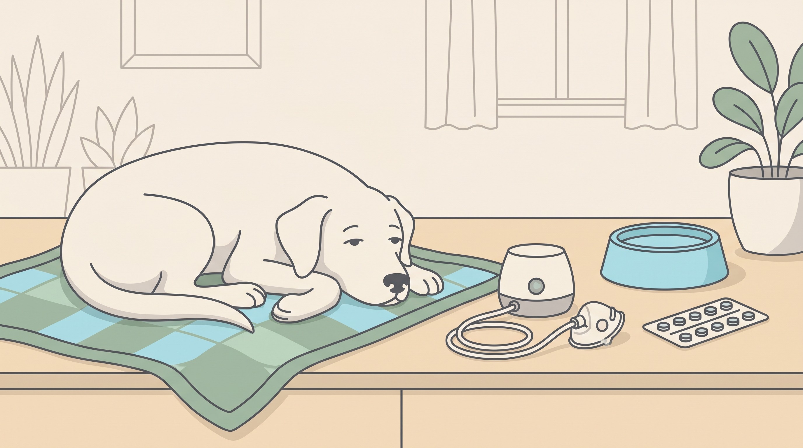
So you've had the diagnosis. Maybe it followed a cough that wouldn't shift, or a moment where your dog was sick and something "went down the wrong way", or an operation, or it landed on top of a laryngeal paralysis you were already managing. Whatever the route in, the word pneumonia has been said, your pet is home with a bag of medication, and the job is now yours. That's a daunting place to be, and I'd like to make it less so.
Here's the reassuring frame first. Most pets treated promptly for pneumonia recover well (American Kennel Club, 2026; MSD Veterinary Manual, n.d.-a). Recovery asks not for heroics but for a handful of small jobs done consistently, plus one steady eye on whether things are heading the right way. That's what this piece is about: the home-care tasks, how to monitor, and how to tell "still recovering, which takes time" from "getting worse, ring the vet now". I'll be honest throughout, including about the bits that are slower and harder than the internet usually admits.
What pneumonia is, briefly
Pneumonia is inflammation deep in the lungs and lower airways that gets in the way of gas exchange, so the blood picks up less oxygen than it should (MSD Veterinary Manual, n.d.-a). That's why the signs you'll have seen all hang together: a deep wet cough, a fever, flatness, going off food, faster or harder breathing, and in the worst cases a bluish tinge to the gums after exertion (MSD Veterinary Manual, n.d.-b).
It usually arrives as a complication rather than out of nowhere. Bacterial pneumonia often follows a viral respiratory infection or is seeded by aspiration, the inhaling of vomit, regurgitated stomach contents, or food or medication that goes the wrong way (MSD Veterinary Manual, n.d.-b). Aspiration runs in well-known company: in one study of 88 dogs the commonest predisposing conditions were oesophageal disease, vomiting, neurological disorders, laryngeal disease and the post-anaesthetic period (Kogan et al., 2008). If your pet fits one of those stories, recovery has a second half to it, which I'll come to. And if you're still piecing together how a cough tipped into this, when a cough becomes pneumonia owns that crossing-the-line moment, so I won't re-tread it here.
The treatment your vet has set, and your part in it
For bacterial pneumonia the backbone is an appropriate antibiotic, ideally chosen with the help of an airway sample sent for culture, with supportive care wrapped around it (Lappin et al., 2017; MSD Veterinary Manual, n.d.-a). Severely affected pets stay in hospital for intravenous antibiotics, oxygen and fluids; mildly affected ones go home on tablets and good nursing (American Kennel Club, 2026). If your pet is home, that's a good sign about where they sit on that scale.
Your single most important job is also the one owners most often get wrong with the best of intentions: finish the entire antibiotic course, exactly as prescribed, even once your pet seems back to normal. Pneumonia clears from the lungs more slowly than it clears from the surface. Traditional guidance was to continue antibiotics until a week after both the clinical signs and the chest x-ray had returned to normal, which often meant several weeks of treatment (MSD Veterinary Manual, n.d.-a). More recent evidence supports shorter, tailored courses for uncomplicated and aspiration pneumonia, sometimes in the region of seven to ten days (Today's Veterinary Practice, n.d.). One study was able to stop antibiotics safely at one to three weeks in most dogs, guided by the pet being clinically well and a normalised inflammatory marker in the blood rather than by a fixed calendar (Fernandes Rodrigues et al., 2022). The length is the vet's call to make, not the cough's, and it never means stopping early off your own bat because the patient looks well. Pneumonia can relapse, and stopping short is one of the surest ways to invite it back.
One safety line I want to be very firm about. Do not give cough suppressants for pneumonia. The cough is hard to listen to, but here it is doing essential work, physically clearing infected material up and out of the lungs, so suppressing it is counterproductive and can do harm (Today's Veterinary Practice, n.d.). This is the opposite of the advice you may have read for a dry chronic cough, where a suppressant can have a proper place. Please don't reach into the cupboard for a leftover medicine or anything made for humans. If the cough is distressing your pet, that's a conversation for your vet, not a problem to solve from the medicine drawer.
The home-care jobs that actually help
Beyond giving the medication faithfully, a few hands-on tasks genuinely speed clearance. Your vet will tell you which apply, so treat this as the menu, not a prescription.
Keep them well hydrated, because here water really is medicine. Your pet's lungs have a built-in cleaning system, a mucociliary "escalator" of tiny hairs that sweeps infected mucus upward and out, and it only works properly when the patient is well hydrated; let them dry out and the secretions thicken and the escalator stalls (Today's Veterinary Practice, n.d.). So fresh water always within reach, gentle encouragement to drink, and in hospital the fluids on the drip, all earn their keep.
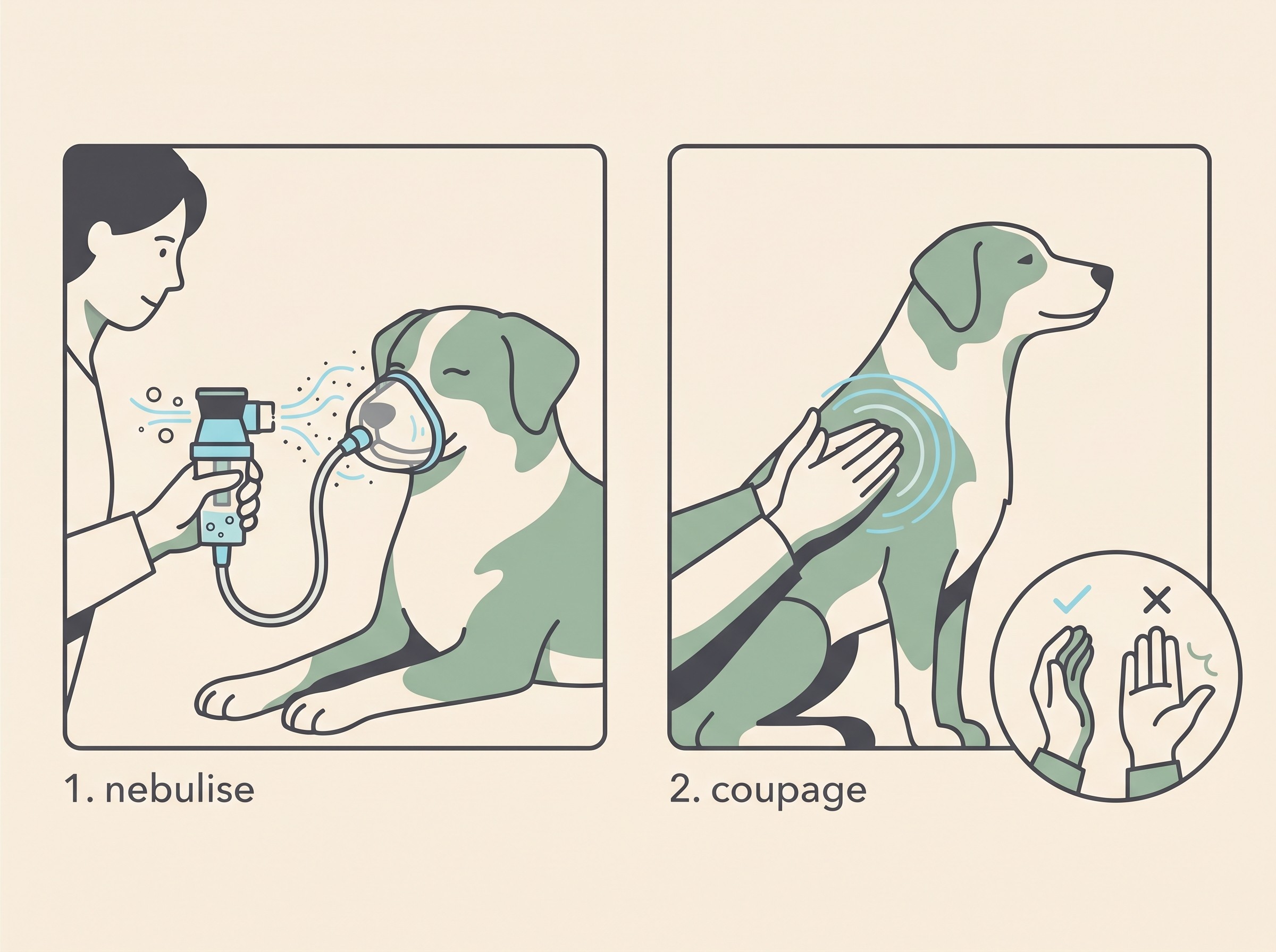
Nebulising, if your vet advises it: a fine saline mist, never steam. Nebulisation pushes a cloud of microscopically fine water droplets deep into the small airways to hydrate and loosen secretions. The catch is that it only works with a proper nebuliser, one producing droplets roughly in the one-to-five-micron range, small enough to travel right down into the lung (VETgirl, n.d.; Veterinary Partner, n.d.). This is the part I most want to land: a steam vaporiser, a kettle, or a room humidifier does not do this job. Their droplets are far too big and never get past the nose and throat, so they do nothing for pneumonia down in the lungs (Veterinary Partner, n.d.). The usual approach is to nebulise a small amount of sterile saline, often with the pet in a covered carrier or wearing a mask, for several minutes at a time (VETgirl, n.d.).
Coupage, the cupped-hand chest clap, straight after nebulising. Coupage means clapping the chest wall firmly but gently with a cupped hand, so air trapped between your palm and their side creates a soft vibration that shakes mucus loose; a flat hand simply doesn't work (VETgirl, n.d.).
Gentle movement, not total stillness. Once your pet is stable, lying motionless all day works against you. Frequent changes of position and short, gentle walks as tolerated help mobilise secretions (Today's Veterinary Practice, n.d.). Aim for movement that helps drainage without the exertion or heat that pushes their breathing into the red. Little and often, on the lead, is the rhythm.
If pneumonia followed an aspiration event, recovery also means lowering the odds of the next one, with changes to how your pet eats and drinks: small frequent meals, the right food consistency, and for some dogs raised or upright feeding, plus steering clear of swimming and great gulps of water (Cornell University, n.d.; Kogan et al., 2008). Dogs with laryngeal paralysis stay aspiration-prone throughout recovery and beyond, even after a tie-back operation, where one large study found pneumonia in roughly a fifth of dogs in the first year and around a third by three to four years (Wilson & Monnet, 2016). That full feeding-and-water protocol deserves its own room, so I'll hand you to aspiration pneumonia risk in laryngeal paralysis for it and keep this piece on recovery itself.
How to monitor recovery at home
Three things tell you which way the wind is blowing: your pet's resting breathing rate, their appetite, and their energy. Taken together, day after day, they are a more honest report card than any single good or bad hour.
The most useful single number is the resting or sleeping respiratory rate. While your pet is properly asleep, count how many breaths they take in a full minute (one rise-and-fall of the chest is one breath; count fifteen seconds and multiply by four if that's easier). Healthy, settled dogs and cats sit comfortably under about 30 breaths a minute (Porciello et al., 2016). A rate steadily above 30, and especially one climbing day on day, is a real early warning that recovery is stalling, particularly alongside a dip in appetite or energy. I'll be straight about where that number comes from: it's best validated in pets with heart disease, and we use it here as a general signal of resting tachypnoea, breathing faster than it should at rest (Porciello et al., 2016). That honesty doesn't weaken it. A rising resting rate is worth a phone call whatever textbook it came from.
This is exactly the kind of trend that's easy to feel and hard to remember, so write it down. The resting respiratory rate tracker turns it into a tap-to-count habit and a line on a graph, and the airway diary lets you log the cough, appetite, energy and breathing rate together so a pattern, good or bad, shows up across the days. A graph drifting gently downward over a week is one of the most reassuring things you can have in front of you. One creeping upward is your cue to act before it becomes a crisis.
When "recovering" has tipped into "getting worse"
Recovery is rarely a straight line, but the overall direction should be the right one. Ring your vet promptly if you see the picture turn: a cough that worsens or comes back after improving, a returning fever, new flatness or lethargy, going off food again, or breathing that gets faster or more laboured, with more effort or heaving from the belly (American Kennel Club, 2026; MSD Veterinary Manual, n.d.-a).
And the line that does not wait: blue, purple or grey gums, or severe struggling to breathe, is an emergency, go now (American Kennel Club, 2026). In a cat, breathing with the mouth open belongs in that same go-now category; cats almost never breathe through an open mouth unless they are in real trouble, so don't wait and don't stop to film it. For the full how-worried-should-I-be sort, is my pet's breathing an emergency owns that triage, so I'll point you there rather than repeat it.
There's also a quieter checkpoint. If there's no improvement after about 48 to 72 hours of treatment, that's when your vet will want to reassess, because it can mean the antibiotic isn't the right match, or there's a complication, or an underlying cause hasn't been dealt with (MSD Veterinary Manual, n.d.-a; Lappin et al., 2017). Not improving on schedule isn't a failure on your part. It's information, and far better acted on early than late.
The honest timeline, and where this is heading
Let me set your expectations truthfully, because false speed only breeds worry. Pneumonia gets better over weeks, not days, and the recovery to feeling fully like themselves can run from about a week to more than a month depending on how poorly they were (American Kennel Club, 2026). The chest x-ray lags behind the patient too, so the lungs can still look hazy on a film after your pet seems their old self, which is precisely why antibiotics carry on past the point they look recovered and why your vet will want a follow-up x-ray to confirm the lungs have genuinely cleared (MSD Veterinary Manual, n.d.-a). That recheck isn't bureaucracy. It's how we make sure treatment stops because the lung is clear, not just because the cough went quiet.
The outlook, on the whole, is encouraging. Reported survival for dogs with aspiration pneumonia sits roughly in the 77 to 88% range, with one large series putting it at about 81.6%, and a feline study reporting around 89% (Kogan et al., 2008; Tart et al., 2010; Today's Veterinary Practice, n.d.). Good but not guaranteed is the fair way to hold it. The pets who do less well are usually those who needed intensive oxygen support, or who have an ongoing underlying cause still driving the problem, such as a progressing laryngeal paralysis (American Kennel Club, 2026; Kogan et al., 2008). That's the real argument for treating the cause, not just the chest.
If you do the unglamorous things well, give every dose, keep the water flowing, nebulise and coupage if you've been asked to, watch that resting rate, and resist the urge to stop early, you've stacked the odds firmly in your pet's favour. Most of recovery happens quietly, on a blanket, while you simply keep the routine going. Keep the resting-rate count ticking over in the tracker as the weeks pass, take the win when the recheck x-ray comes back clear, and let that downward graph be the proof you're allowed to relax.
References
- American Kennel Club. (2026). Aspiration Pneumonia in Dogs (G. Johnstone, with veterinary input from A. Odunayo & A. Smith). Retrieved from
- Cornell University, Riney Canine Health Center. (n.d.). Laryngeal Paralysis. Retrieved from
- Fernandes Rodrigues, N., Giraud, L., Bolen, G., Fastrès, A., Clercx, C., Boahen, F., Idziorek, S., & Merveille, A.-C. (2022). Antimicrobial discontinuation in dogs with acute aspiration pneumonia based on clinical improvement and normalization of C-reactive protein concentration. Journal of Veterinary Internal Medicine, 36(3), 1082–1088.
- Kogan, D. A., Johnson, L. R., Sturges, B. K., Jandrey, K. E., & Pollard, R. E. (2008). Etiology and clinical outcome in dogs with aspiration pneumonia: 88 cases (2004–2006). Journal of the American Veterinary Medical Association, 233(11), 1748–1755.
- Lappin, M. R., Blondeau, J., Boothe, D., Breitschwerdt, E. B., Guardabassi, L., Lloyd, D. H., Papich, M. G., Rankin, S. C., Sykes, J. E., Turnidge, J., & Weese, J. S. (2017). Antimicrobial use guidelines for treatment of respiratory tract disease in dogs and cats: Antimicrobial Guidelines Working Group of the International Society for Companion Animal Infectious Diseases. Journal of Veterinary Internal Medicine, 31(2), 279–294.
- MSD Veterinary Manual. (n.d.-a). Pneumonia in Dogs and Cats (professional version). Retrieved from
- MSD Veterinary Manual. (n.d.-b). Pneumonia in Dogs (pet-owner version). Retrieved from
- Porciello, F., Rishniw, M., Ljungvall, I., Ferasin, L., Häggström, J., & Ohad, D. G. (2016). Sleeping and resting respiratory rates in dogs and cats with medically-controlled left-sided congestive heart failure. The Veterinary Journal, 207, 164–168.
- Tart, K. M., Babski, D. M., & Lee, J. A. (2010). Potential risks, prognostic indicators, and diagnostic and treatment modalities affecting survival in dogs with presumptive aspiration pneumonia: 125 cases (2005–2008). Journal of Veterinary Emergency and Critical Care, 20(3), 319–329.
- Today's Veterinary Practice. (n.d.). Treating Bacterial Pneumonia in Dogs and Cats. Retrieved from
- Veterinary Partner (VIN). (n.d.). Nebulizer Use for Dogs and Cats. Retrieved from
- VETgirl. (n.d.). How to Nebulize and Coupage a Dog With Aspiration Pneumonia. Retrieved from
- Wilson, D., & Monnet, E. (2016). Risk factors for the development of aspiration pneumonia after unilateral arytenoid lateralization in dogs with laryngeal paralysis: 232 cases (1987–2012). Journal of the American Veterinary Medical Association, 248(2), 188–194.
Keep track of how your pet is doing
The owners who cope best are the ones who notice changes early. A simple health log shows you what is working, and what is not, before the next vet visit.
Start tracking, freeYou're not doing this alone
Compare treatment journeys and talk to owners managing breathing & airways. Free to join.
Join PetsLikeMine