IVDD in Dogs: What It Is and Why Discs Go Wrong
Claire Greenway
BVM&S MRCVS
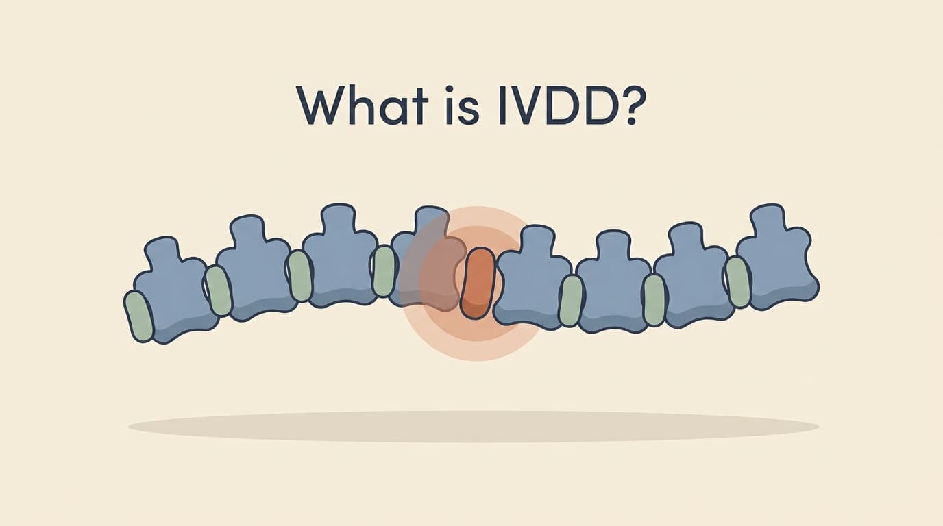
IVDD, intervertebral disc disease, is a common spinal condition in dogs, and the letters alone can be unsettling when a vet first mentions them, especially since a quick search online tends to throw up the frightening end of the spectrum first. It comes in several forms ranging from mild to serious, and understanding what it actually is takes a good deal of the fear out of it. This guide is orientation, not emergency advice: a plain-English explanation of what the discs are, why they go wrong, and the main types, so the rest of the picture makes sense. If you are in the middle of a crisis right now, our guide to the red flags and our triage checker are the place to start instead.
What a disc is, and what it does
To understand the disease, it helps to picture the healthy structure first. Your dog's spine is a chain of bones, the vertebrae, running from the neck to the tail, and between most pairs of vertebrae sits an intervertebral disc. These discs are the spine's cushions, the shock absorbers that sit between the bones, letting the back bend, flex, and twist while softening the everyday jolts of running and jumping.
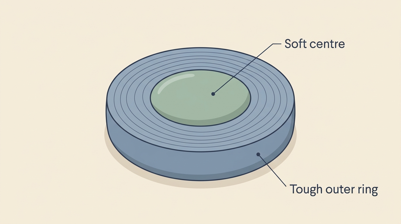
A useful way to picture a single disc is as a small jam doughnut. It has a soft, jelly-like centre, called the nucleus pulposus, which is mostly water held in place by special proteins, and a tougher, fibrous outer ring that contains that centre, called the annulus fibrosus. When everything is healthy, the springy, water-rich centre is safely contained within its outer casing, and the disc does its cushioning job quietly, year after year. The trouble in IVDD comes when that arrangement breaks down, and where it breaks down matters: the nucleus sits slightly toward the top of the disc, nearest the spinal cord running just above, so when a disc fails it tends to push upward, straight toward the cord. That simple piece of geometry is why a disc problem so often becomes a spinal-cord problem.
How a disc fails
Discs go wrong through a process of degeneration that then leads to herniation, and it is worth understanding the two steps, because they explain almost everything that follows. The first step is degeneration, and at its root is water. The soft centre of the disc depends on special water-holding proteins to stay plump and springy, and these are produced by particular cells within the disc. As a disc degenerates, it loses those cells and proteins, dries out, and stiffens, sometimes hardening and calcifying until it is brittle rather than springy. A degenerated disc is a disc primed for trouble, because it no longer cushions properly and its contents are no longer safely contained.
The second step is herniation, when the damaged disc material pushes out of place. Because of that weak upper edge and the dorsally-placed nucleus, the material tends to herniate upward into the spinal canal, where it presses on, or strikes, the spinal cord. This is the moment that produces the signs owners notice, because a compressed or bruised spinal cord cannot send its messages properly. Depending on how much material comes out, how fast, and how hard it hits the cord, the result ranges from back pain alone, through wobbliness and weakness, to an inability to walk. The spinal cord is delicate and does not tolerate pressure or impact well, which is why a disc problem can have such marked effects, and why the speed and severity of signs matter so much, as our guides to the grades and the red flags explain. With that two-step picture in mind, degeneration then herniation, the different types of IVDD become much easier to follow.
Three patterns to know
IVDD is not a single thing, and knowing which type your dog has makes an enormous difference to what it means. There are three main patterns to know about, and they behave very differently. The classification goes back to a Danish researcher, Hansen, who in the 1950s described the first two, and they are still called Hansen type I and type II today.
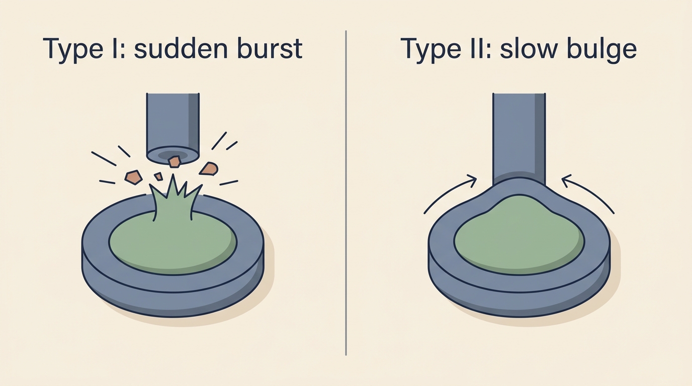
Hansen Type I: the acute "burst" disc
Hansen Type I is the classic, dramatic form, and the one most people picture when they hear IVDD. Here, a disc whose centre has dried out and calcified suddenly ruptures, with the hardened inner material bursting out and extruding into the spinal canal, striking and compressing the cord. The failure can be genuinely catastrophic and sudden, which is why a dog that was fine in the morning can be in pain or unable to walk by the afternoon, and why this type so often constitutes a spinal emergency.
Type I is strongly associated with the chondrodystrophic breeds, the short-legged dogs such as dachshunds, French bulldogs, beagles, and corgis, and there is a real biological reason for that. These breeds are genetically short of the water-holding disc cells from early in life, so their discs degenerate and calcify years ahead of other dogs, often becoming prone to bursting at just three to six years old. Our guides on dachshunds and on the other at-risk breeds go into why these dogs are so predisposed. One important modern clarification, though, since older sources state it too rigidly: Type I extrusions are not exclusive to short-legged breeds. We now know that all dogs undergo this kind of disc degeneration eventually, just more slowly in others, so a larger or longer-legged dog can occasionally have a Type I extrusion too. So if you have one of the classic at-risk breeds, Type I is very much the form to understand and to know the warning signs for, but it is not unheard of in other dogs.
Hansen Type II: the slow "bulge"
Hansen Type II is the quieter, more gradual form. Instead of a sudden burst, the tough outer ring of the disc slowly thickens and bulges, pressing on the spinal cord progressively over months. Because it comes on slowly, the signs creep in rather than appearing overnight, often a gradual stiffness, weakness, or wobbliness that worsens over time, and because the change is so gradual, Type II disc bulges are quite often found incidentally and may cause few or no signs at all. Type II is seen more in older dogs and in the larger, non-chondrodystrophic breeds such as Labradors and German Shepherds, usually in dogs over about seven years old. It is a different beast from Type I, slower, more insidious, and managed differently, though the underlying idea, disc material pressing on the cord, is the same.
Type III (ANNPE): the traumatic "missile" disc
The third type is worth explaining properly, because it is widely misunderstood and the truth about it is genuinely reassuring. Type III, often called ANNPE, stands for acute non-compressive nucleus pulposus extrusion, and it is sometimes nicknamed a "missile disc." Here, a relatively healthy disc, under a sudden burst of force during vigorous exercise or play, fires a small amount of its soft centre out at high velocity, like a pellet. The crucial difference from Type I is in the name: it is non-compressive. The material strikes and bruises the spinal cord as it shoots past, causing sudden signs, but then it is gone, so unlike Type I there is no lump of material left behind pressing on the cord.
This changes everything about the outlook. Because there is nothing left compressing the cord, ANNPE usually does not require surgery, and the treatment is rest and rehabilitation while the bruise to the cord heals. And the prognosis is frequently good, with many dogs recovering well over time, since the injury is often a one-off bruise rather than ongoing damage. So while an ANNPE can look frightening at the moment it happens, a sudden yelp and stumble or collapse mid-run, often with no apparent pain afterwards, it is often one of the more hopeful forms of disc injury, and understanding that it exists, and that it is non-compressive and usually non-surgical, can be a real comfort to an owner told their dog has had this kind of disc event. It is a reminder that not every disc injury is the dreaded burst disc, and not every one needs surgery.
How common is it, and is it always serious?
It helps to end with a sense of proportion, because the word IVDD covers a huge range. Disc degeneration itself is extremely common as dogs age: by the time they reach middle age and beyond, a large proportion of dogs show some disc degeneration when it is looked for closely, and a smaller number have a disc actually bulging. The great majority of that never causes a serious problem. So having "disc disease" mentioned does not automatically mean a catastrophe, in many dogs it means a slow, manageable, or even silent change, and only a minority go on to the dramatic, acute events that make IVDD frightening.
That said, when an acute Type I extrusion does happen, it can be serious and time-sensitive, which is exactly why knowing the warning signs matters so much for the at-risk breeds. The reassuring overall picture is one of a spectrum: at one end, the common, mild, and often incidental disc changes of an ageing spine; at the other, the uncommon but urgent burst disc that needs prompt attention. Knowing where on that spectrum your own dog sits is what the rest of our guides help you work out.
So, that is the foundation: a disc is a water-filled cushion with a soft centre and a tough ring, IVDD is what happens when that centre dries out and the disc fails and presses on the spinal cord, and the type, a sudden Type I burst, a slow Type II bulge, or a traumatic but non-compressive Type III, shapes what it means and how it is treated. From here, the rest of the picture follows naturally: if you want to know how serious a given case is, our guide to the grades explains how vets measure severity and what the recovery odds are, and if you are worried your dog is showing signs right now, our guide to the red flags and our triage checker will help you judge how urgently to act.
References
- Olby NJ, Moore SA, Brisson B, Fenn J, Flegel T, Kortz G, Lewis M, Tipold A. ACVIM consensus statement on diagnosis and management of acute canine thoracolumbar intervertebral disc extrusion. Journal of Veterinary Internal Medicine, 2022;36(5):1570-1596.
- Demographic and lifestyle characteristics impact lifetime prevalence of owner-reported intervertebral disc disease: 43,517 companion dogs in the United States. Journal of the American Veterinary Medical Association, 2025;263(5).
- Rusbridge C. Canine chondrodystrophic intervertebral disc disease (Hansen type I disc disease). BMC Musculoskeletal Disorders, 2015;16(Suppl 1):S11.
- Brown EA, Dickinson PJ, Mansour T, et al. FGF4 retrogene on CFA12 is responsible for chondrodystrophy and intervertebral disc disease in dogs. Proceedings of the National Academy of Sciences, 2017;114(43):11476-11481.
- Togawa G, Lewis MJ, Devathasan D, et al. Outcome of conservative and surgical management in dogs with acute non-compressive nucleus pulposus extrusion and fibrocartilaginous embolic myelopathy. Frontiers in Veterinary Science, 2024;11:1406843.
Free downloads
Companion worksheets to put what you've read into practice. Free PDFs, print at home.

Sister tool · Sightline
Track whether treatment is working
Sightline, a separate ConciergeVet tool, runs a short weekly check-in built on validated pain and mobility instruments, turns it into a single Sightline Score you can watch trend over weeks rather than judge on one bad day, and produces a report you can bring to your vet.
A written log, or our printable trackers, does much the same job.
See how Sightline tracks treatmentTrack the recovery, day by day
Recovery from a disc injury is all about the trend. Log the daily milestones through crate rest and rehab so you can see real progress, not just good and bad days.
Open the Recovery TrackerYou're not doing this alone
Compare treatment journeys and talk to owners managing IVDD. Free to join.
Join PetsLikeMine