How Arthritis Is Diagnosed: Understanding the Vet Visit
Claire Greenway
BVM&S MRCVS
Most owners notice the same odd thing. The dog who was stiff, slow and reluctant at home walks into the consulting room and turns into a bright-eyed, bouncy version of themselves on the floor. Adrenaline and unfamiliar surroundings mask a remarkable amount of discomfort, so by the time the vet is examining them, your dog often looks perfectly fine.
That is one of the reasons arthritis diagnosis can feel frustrating. You know something has changed at home, but the evidence isn't on display when you need it to be. Your vet is trying to piece together a picture from what they can see in fifteen minutes, what they can feel during examination, and what you can tell them about the rest of your dog's life.
So this article explains how that diagnostic process actually works, what your vet is looking for, what tests might be involved, and what the results actually mean. The aim is to give you a thorough understanding of what's happening so that you can be an active participant in the process rather than a confused bystander.
What your vet is actually doing in the consultation
A good arthritis consultation should never be rushed. If your appointment feels squeezed into ten minutes between other cases, it's worth asking for a longer slot or a dedicated orthopaedic appointment. Proper assessment of an arthritic dog takes time.
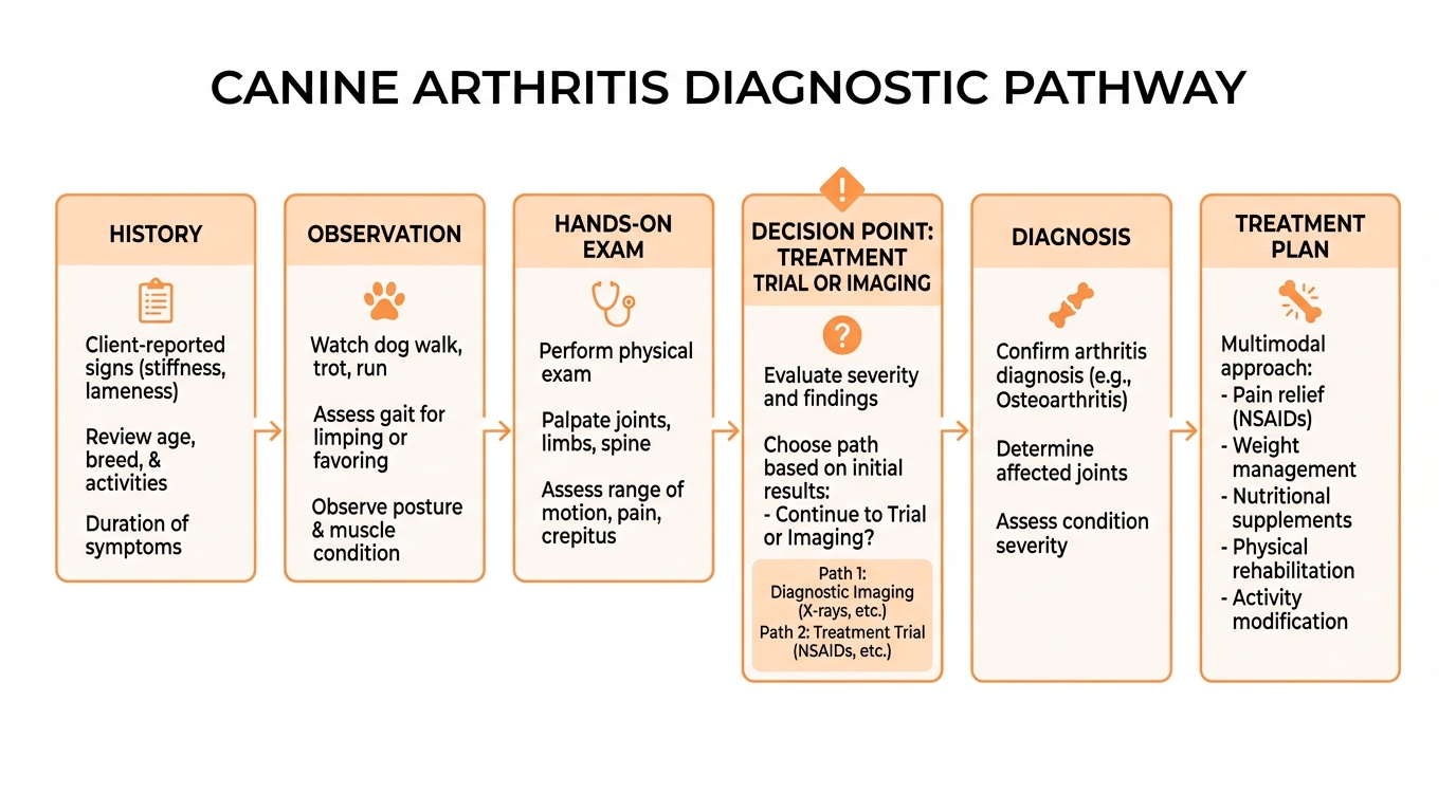
A consultation typically has three main parts: the history, the observation and the hands-on examination. Each one gives different information, and a confident diagnosis usually requires all three to point in the same direction.
Starting with the history
This is where you come in. Your observations at home are not just helpful, they're often the single most important diagnostic tool. A vet who skips this stage and goes straight to palpating joints is missing the most valuable information available.
Expect to be asked about onset (when did you first notice something?), progression (has it got worse, stayed the same, or fluctuated?), specific signs (limping, stiffness, behavioural changes, reluctance to exercise), what makes things better or worse (warmth, rest, exercise, weather), and what your dog can no longer do that they used to. Your vet will also want to know about your dog's general health, any previous injuries or surgeries, medications they're currently on, and their typical exercise routine.
If you've kept the observation diary I recommend in our article on spotting the signs, this is where it pays off. Hand it over, or work through it together. A clear, specific history dramatically changes what your vet can conclude.
Watching how your dog moves
Before your vet lays a hand on your dog, they should be watching. How does your dog stand? Are they shifting weight off any particular leg? Is their posture even, or are they slightly hunched, or favouring one side?
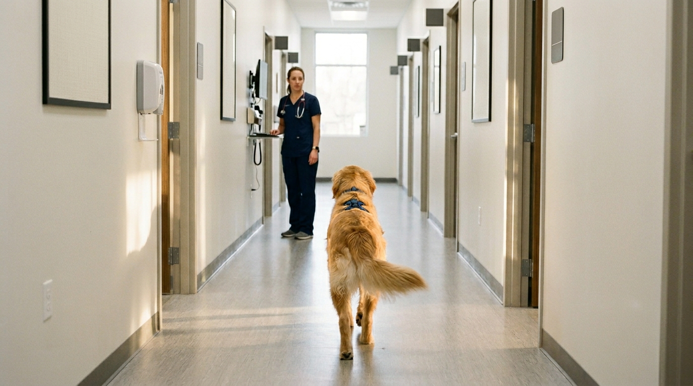
If your consulting room is big enough, your vet may ask you to walk your dog away and back across the room, then turn. Subtle asymmetries in gait are often visible at a walk but disappear at a trot, or vice versa. Some vets will ask to take your dog into the corridor or car park for a slightly longer assessment, which can be much more revealing than what you can see in a six-metre consulting room.
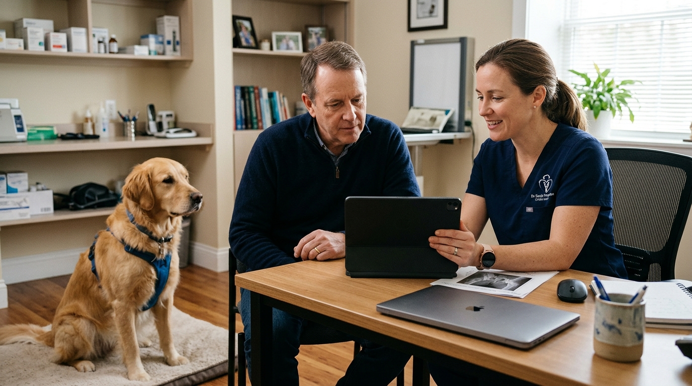
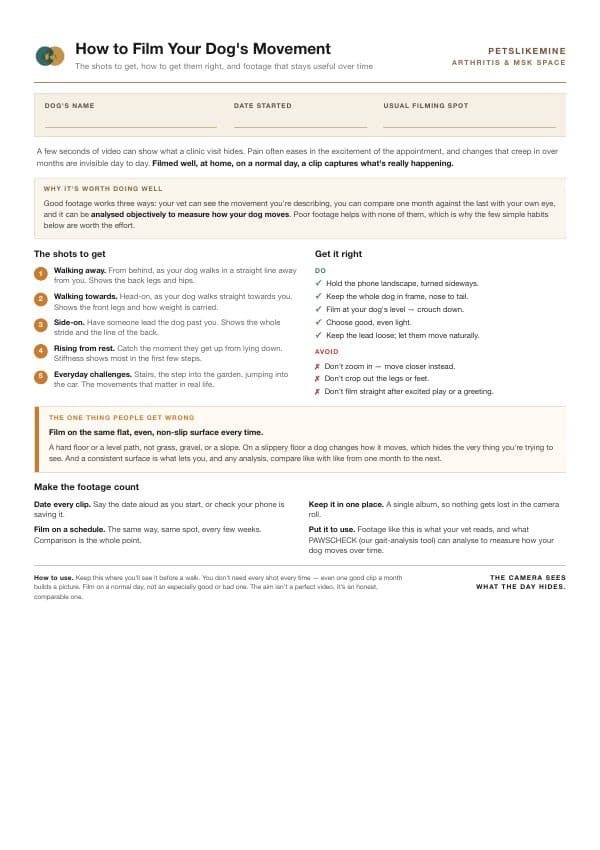
One important note: if your dog is anxious or excited in the clinic, the observation phase often reveals very little. This is where pre-recorded video at home becomes invaluable. A 30-second clip of your dog rising from rest, walking down a hallway, or going up stairs at home tells your vet more than several minutes of clinical observation in many cases. If you have video, show it. That same home footage can also be analysed objectively, complementing the vet's hands-on assessment and giving a measured baseline to compare against later.
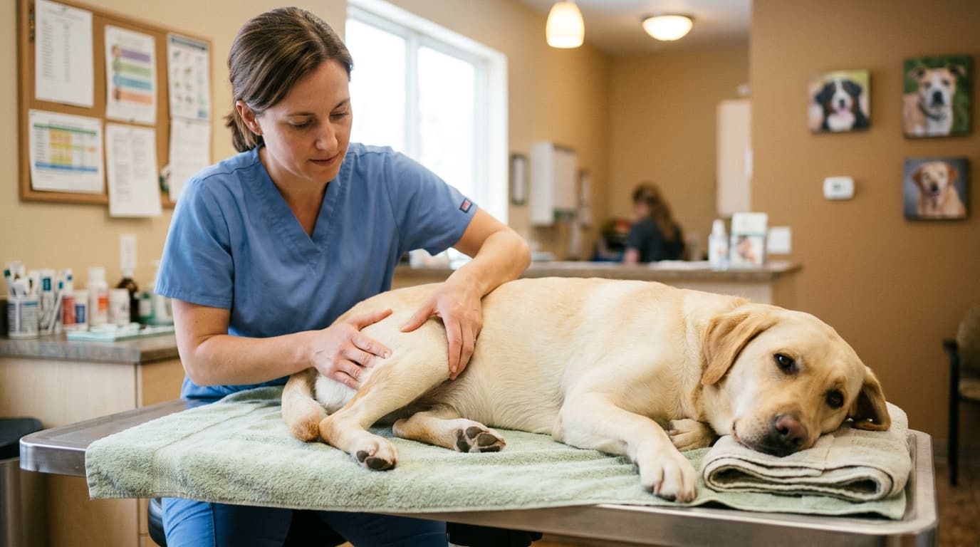
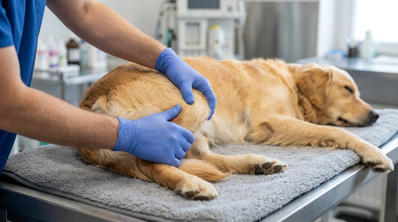
The hands-on examination
This is the part most owners think of as "the examination," but as you can see, it should be the third stage of a longer process.
A proper orthopaedic examination involves systematically working through each joint in turn, usually starting at the front and working backwards, or starting at the painful area and moving outward. For each joint, your vet is assessing:
Range of motion. Can the joint be flexed and extended through its normal range without resistance? An arthritic joint often has a reduced range of motion compared to a healthy one. Your vet will compare left to right where possible.
Pain on manipulation. Does flexing, extending, or rotating the joint produce a pain response? This might be obvious (a yelp, a snap, pulling the leg away) or very subtle (lip licking, head turning, slight body tension).
Crepitus. A grinding, crunching, or crackling sensation felt through the joint as it moves. Crepitus indicates roughened joint surfaces and is a fairly reliable sign of structural arthritis.
Joint swelling or effusion. A normal joint has a sharp, defined outline. An arthritic joint often feels thickened or "fuller" due to thickening of the joint capsule and sometimes increased synovial fluid. Some joints are easy to assess this way (the stifle, for example). Others are harder.
Muscle mass. A leg that has been used less due to chronic pain often shows muscle atrophy. Your vet will compare left to right, looking for asymmetric muscle loss that suggests one side has been favoured.
Specific orthopaedic tests. For certain joints, there are specific manipulations that test for particular conditions. The cranial drawer test and tibial thrust test check for cruciate ligament instability in the stifle. The Ortolani test assesses hip laxity. Manipulation of the elbow can reveal specific patterns of dysplasia. These tests aren't always done in a routine consultation, but a vet who suspects a specific condition will use them.
A thorough orthopaedic examination of a moderately sized dog with multi-joint problems can easily take 10 to 15 minutes on its own. If your vet has flown through it in two minutes, the examination has been superficial. That's not necessarily wrong for a quick reassessment, but for an initial workup of suspected arthritis, it's not enough.
What your vet might suggest next
After history, observation, and examination, your vet will often have a working diagnosis. They may say "this looks consistent with osteoarthritis, probably most significant in the hips and elbows." Or they may say "there's clearly some discomfort but I want imaging before I commit to a diagnosis." What happens next depends on your dog, on what they've found, and on the resources available.
Trying treatment first
In some cases, particularly with older dogs showing classic signs, your vet may suggest starting treatment without further investigation. This is sometimes called a "therapeutic trial." If your dog responds well to a short course of anti-inflammatory medication, that response itself becomes part of the diagnosis. If the medication doesn't help, that's also useful information that prompts further investigation.
This approach is reasonable when the clinical picture is consistent, the dog is otherwise healthy, and the cost of imaging would significantly affect the family's ability to fund ongoing treatment. It's less appropriate for young dogs (where you really want to know what's structurally going on), dogs with concurrent illness, or when the picture is atypical.
Blood tests
Blood tests don't diagnose osteoarthritis directly, but they're often done before starting long-term anti-inflammatory medication. The goal is to establish a baseline for kidney and liver function, since these are the organs that process most of the drugs we'd be using. Repeat blood tests at intervals (typically before starting and then 4-6 weeks after, with periodic checks every 6 to 12 months) help monitor for side effects of long-term medication.
In some cases, blood tests are also used to rule out other conditions that can mimic arthritis. Tick-borne diseases like Lyme disease and Anaplasma can cause joint pain and lethargy. Endocrine conditions like Cushing's disease and hypothyroidism can affect muscle and joint comfort. If your dog has atypical signs, a broader blood screen may be worth doing.
Expect to pay somewhere between £80 and £200 for a comprehensive blood panel in UK first-opinion practice.
Radiographs (X-rays)
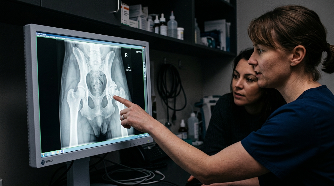
Radiographs are the most commonly used imaging modality for diagnosing osteoarthritis. They're widely available, relatively affordable, and very useful for visualising structural joint change.
What X-rays show in arthritis:
- Osteophytes (new bone growth around the joint margins, often called "bone spurs")
- Subchondral sclerosis (increased density of the bone immediately beneath the cartilage)
- Joint space narrowing (in advanced cases)
- Subchondral cysts
- Soft tissue swelling
- Changes consistent with specific underlying conditions (hip dysplasia, elbow incongruity, OCD lesions)
What X-rays don't show:
- The cartilage itself (cartilage isn't visible on X-ray)
- Soft tissue structures like ligaments and tendons
- The degree of pain your dog is experiencing
That last point is important. The correlation between X-ray findings and clinical pain is genuinely poor. We regularly see dogs with dramatic radiographic changes who function reasonably well, and dogs with relatively modest X-ray findings who are in significant pain. X-rays tell us about structural disease. They don't tell us about pain experience. Both pieces of information matter, but neither replaces the other.
To get good diagnostic radiographs, your dog usually needs to be sedated. Conscious radiographs are sometimes possible for cooperative dogs, but for proper positioning of joints (particularly hips and elbows) sedation produces much better quality images. Sedation also allows for a more thorough orthopaedic examination at the same time, since a relaxed dog is much easier to assess.
Expect to pay £200 to £600 for radiographs in UK first-opinion practice, depending on the number of views and whether sedation is included. Referral imaging will typically be at the higher end.
CT (Computed Tomography)
CT scans provide cross-sectional images of bony structures in much more detail than plain radiographs. They're particularly useful for:
- Complex joint conditions where the geometry is hard to assess on plain films (elbow dysplasia is a classic example)
- Pre-surgical planning for orthopaedic procedures
- Detecting subtle bony lesions that aren't visible on standard radiographs
- Investigating spinal disease
CT requires general anaesthesia for dogs, since they need to lie completely still in a precise position. The scan itself is quick (often just a few minutes), but the overall visit involves anaesthesia, recovery, and discharge.
UK CT scans for orthopaedic indications typically cost £600 to £1,500, depending on the area scanned, the facility, and whether it's part of a wider diagnostic workup. CT is usually performed at referral centres or specialist imaging facilities rather than in general practice.
MRI (Magnetic Resonance Imaging)
MRI is the best modality for visualising soft tissue structures like cartilage, ligaments, tendons, and the spinal cord. For pure osteoarthritis it's rarely necessary, since the bony changes show up well on X-ray or CT. But MRI becomes important when:
- Soft tissue injury is suspected alongside or instead of arthritis
- Spinal disease is being investigated
- Surgical planning requires detailed soft tissue information
- The cause of pain isn't explained by the bony findings
MRI requires general anaesthesia and significantly more time than CT (a scan typically takes 45 to 90 minutes). UK MRI for dogs typically costs £1,500 to £2,500.
Arthroscopy
For some conditions, particularly elbow dysplasia and some cruciate problems, the most informative diagnostic test is arthroscopy. This is a keyhole examination of the joint using a small camera, performed under general anaesthesia. It allows direct visualisation of the joint surfaces and is often combined with treatment in the same procedure (removing loose fragments, flushing the joint, and so on).
Arthroscopy is usually performed by orthopaedic specialists and is typically used when other diagnostics have suggested a specific problem that needs further definition or treatment. Costs vary widely depending on the joint and what's done, but expect £1,500 to £3,500 for arthroscopic procedures at referral centres.
Joint fluid analysis
In some cases, particularly when the cause of joint pain is unclear or when an inflammatory or infectious arthritis is suspected, your vet may want to sample the joint fluid. A needle is inserted into the joint (under sedation or anaesthesia) and a small amount of synovial fluid is withdrawn for analysis. This can distinguish osteoarthritis from immune-mediated polyarthritis or septic arthritis, which require very different treatment.
Decoding your dog's report
When you get the results of imaging, the language can be intimidating. Here's a translation of some of the terms you might encounter.
"Mild/moderate/severe degenerative joint disease (DJD)." This is the same as osteoarthritis. Mild generally means a few small osteophytes and minimal joint changes. Moderate means more extensive changes with clear evidence of disease. Severe means significant joint remodelling, large osteophytes, and often joint space changes.
"Osteophytosis" or "osteophyte formation." New bone growth around the joint margins. This is one of the hallmarks of osteoarthritis. The location and extent matter more than the simple presence of osteophytes.
"Subchondral sclerosis." Increased density of the bone immediately beneath the joint surface. This represents the bone trying to compensate for damaged cartilage above it. It's a sign of moderate to advanced disease.
"Joint effusion." Increased fluid within the joint. This indicates active inflammation. It's commonly seen in arthritis but can also indicate other joint problems.
"Bilateral changes." The same condition is present on both sides. In arthritis this is very common, particularly for conditions like hip dysplasia or elbow dysplasia.
"Hip dysplasia, OFA grade X" or "BVA hip score of X/53." Specific scoring systems for hip conformation. Lower numbers are better. These scores are used both diagnostically and for breeding decisions.
"Elbow dysplasia" is an umbrella term covering several specific conditions: fragmented coronoid process, ununited anconeal process, osteochondritis dissecans, and elbow incongruity. The specific subtype affects treatment options.
"Spondylosis deformans" is bony bridging between the vertebrae of the spine. It's often visible on X-rays of older dogs and is sometimes incidental, but can contribute to pain and stiffness.
"Cranial cruciate ligament disease" is the most common cause of stifle (knee) arthritis in dogs. The ligament fails progressively, leading to instability and secondary arthritis.
"Within normal limits" or "no significant findings" means nothing dramatic was seen on the imaging. This doesn't necessarily mean there's no arthritis (early disease may not show clearly on radiographs), but it suggests the structural changes aren't severe.
If you don't understand something in your dog's report, ask. A good vet will go through the findings with you and explain what they mean for management. If you feel rushed, ask for a follow-up appointment specifically to discuss the results.
What if the diagnosis isn't clear?
Sometimes a thorough workup doesn't produce a definitive answer. The X-rays show some changes but not enough to explain the pain. The orthopaedic examination is suggestive but not conclusive. Your dog responds partially to a therapeutic trial but not fully.
This is more common than you might think. Several conditions can mimic or coexist with arthritis:
Soft tissue injuries like partial cruciate tears, biceps tendinopathy, or iliopsoas strain can cause similar signs to arthritis but require different management.
Lumbosacral disease can cause pain that looks like hip arthritis but is actually originating from the lower spine.
Polyarthritis (immune-mediated joint disease) can present with multi-joint pain but requires very different treatment from osteoarthritis.
Neurological conditions like degenerative myelopathy can cause hindlimb weakness that mimics hip arthritis.
Cancer of the bone or surrounding tissues can occasionally present similarly to arthritis, particularly in larger dogs.
If your dog's diagnosis isn't clear, or if treatment isn't producing the expected response, don't be afraid to ask about referral to a specialist. Orthopaedic surgeons and rehabilitation specialists see these complex cases regularly and have access to diagnostic tools that aren't available in general practice.
When to ask for referral
Most arthritis can be diagnosed and managed perfectly well in first-opinion practice. But there are situations where specialist input adds significant value:
- Your dog is young (under five) with significant orthopaedic disease, where surgical options might change the long-term outcome
- The diagnosis isn't clear after initial workup
- Standard medical management isn't working
- Surgery is being considered
- You want a second opinion before committing to a long-term management plan
- Your dog has multi-joint disease that is difficult to manage
- There are complicating factors (heart disease, kidney disease, and so on) that make medication choices difficult
Asking for referral isn't a criticism of your vet. Good vets refer cases they think will benefit from specialist input, and many will recommend it themselves. If you raise it and your vet seems defensive, that's a warning sign in itself.
Specialist referral typically requires a letter of referral and the imaging from your first-opinion vet. Initial consultations at referral centres typically cost £200 to £400, with further investigations and treatment costed separately.
What to expect emotionally
A few practical thoughts on the diagnostic process from the perspective of someone who has guided many owners through it.
It's normal to feel overwhelmed. Even a relatively simple arthritis diagnosis can come with a lot of new information all at once. Medications, costs, future implications, lifestyle changes. It's a lot. Take notes. Ask for written information. Book a follow-up appointment to revisit the discussion once you've had time to absorb things.
It's normal to feel guilty. Many owners come away from a diagnostic appointment thinking "I should have noticed sooner" or "I shouldn't have let her exercise so much." Please let that go. You're noticing now, and that's what matters. The signs are designed to be missed.
It's normal to feel relieved. Some owners find that having a name for what's been going on actually helps. The vague worry about "what's wrong with my dog" becomes a specific condition with specific management options. This is often a positive emotional shift.
It's normal to ask for time. If your vet is recommending a treatment plan or further investigations, you don't have to commit on the spot. "Can I think about this and come back to you?" is a perfectly reasonable response. Pressure to make decisions immediately isn't usually warranted in chronic disease management.
It's normal to ask about cost. Veterinary care in the UK varies widely in price, and it's reasonable to ask about the cost of each step before agreeing to it. A good vet will discuss this openly and help you find the best path for your situation. If money is a constraint, say so. Many things can be modified to fit different budgets.
You've already done the hard part
If you've got to the point of bringing your dog in for an arthritis assessment, you've done the hard part. You've noticed the changes, you've taken them seriously, and you've sought help. The diagnostic process from here is largely a matter of working through it with your vet to understand what's going on and what to do about it.
Whatever the specific diagnosis, you're now in a much better position than you were before. You know what you're dealing with. You can make informed decisions. You can advocate effectively for your dog.
References
- Gordon WJ, Conzemius MG, Riedesel E, et al. The relationship between limb function and radiographic osteoarthrosis in dogs with stifle osteoarthrosis. Veterinary Surgery, 2003.
- Moving beyond the limits of detection: the past, the present, and the future of diagnostic imaging in canine osteoarthritis. Frontiers in Veterinary Science, 2022.
Free downloads
Companion worksheets to put what you've read into practice. Free PDFs, print at home.
Sister tool · Sightline
Track whether treatment is working
Sightline, a separate ConciergeVet tool, runs a short weekly check-in built on validated pain and mobility instruments, turns it into a single Sightline Score you can watch trend over weeks rather than judge on one bad day, and produces a report you can bring to your vet.
A written log, or our printable trackers, does much the same job.
See how Sightline tracks treatmentSee where your pet stands today
Tracking is the other half of managing arthritis. Take the 2-minute Mobility Check to see your pet's stage, then watch it shift as treatment takes effect.
Take the Mobility CheckYou're not doing this alone
Compare treatment journeys and talk to owners managing arthritis. Free to join.
Join PetsLikeMine