
Elbow Dysplasia: Understanding the Condition and Its Surgical Options
Dr. Alastair Greenway
MRCVS, 25 years clinical experience
Elbow dysplasia is the most common cause of front-leg lameness in young large-breed dogs, and one of the leading developmental causes of elbow arthritis. If your dog has been diagnosed with it, you may already have run into something that confuses many owners: elbow dysplasia isn't one single thing. It's an umbrella term covering several related developmental problems of the elbow, which is part of why the conversation about it can feel complicated.
This is the third of our condition-specific surgical guides, sitting beneath the general article on the surgery decision, which I'd recommend reading alongside it. Here we go deep on elbow dysplasia specifically: what it actually is, how it's diagnosed, what the options are, and what to expect.
As with all these guides, the aim is to help you understand the condition and have a well-informed conversation with your vet or surgeon, not to tell you which treatment your dog should have. Elbow dysplasia is a genuinely complex area where the right approach depends on exactly which problem your dog has and how severe it is, and that's a judgement for a specialist who has assessed your individual dog. My job is to make you a good partner in that conversation.
What we mean by elbow dysplasia
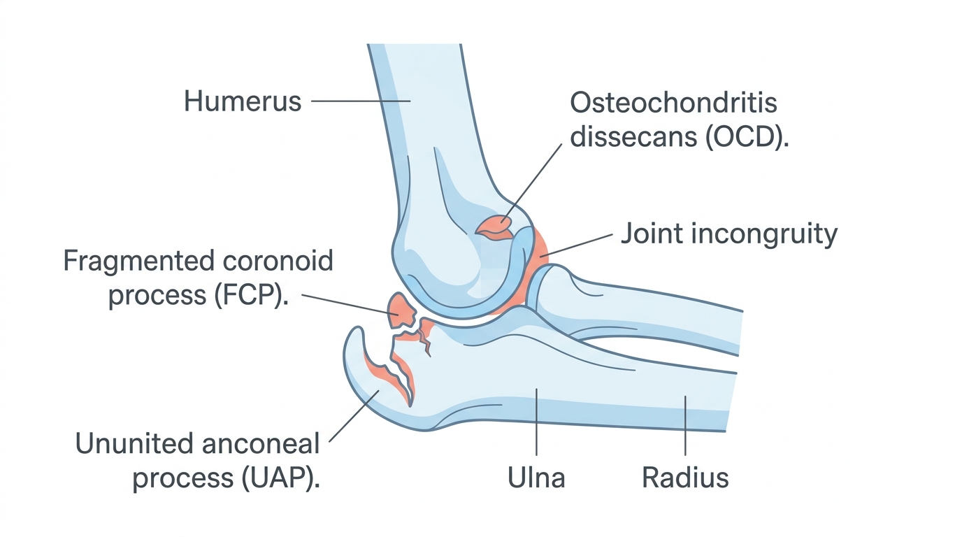
The elbow is a hinge joint made up of three bones, the humerus of the upper arm and the radius and ulna of the forearm, which must fit together with considerable precision. Elbow dysplasia is what happens when, during growth, those bones don't develop or fit together quite right. The resulting imperfect fit, called incongruity, concentrates abnormal forces on small areas of the joint, which causes damage, pain, and arthritis.
Why call it an umbrella term? Because this faulty development shows up in several specific forms, and a dog usually has one of them, occasionally more. The main ones are:
- Medial coronoid disease, including a fragmented coronoid process (FCP), where one of the small bony projections on the ulna cracks or fragments. This is the most common form by far, accounting for the majority of cases.
- Ununited anconeal process (UAP), where another bony projection in the joint fails to fuse properly during growth.
- Osteochondritis dissecans (OCD), where a flap of joint cartilage fails to form properly and separates.
- Joint incongruity, where the bones simply don't fit together evenly.
These can occur alone or in combination, and the medial compartment, the inner part of the joint, is where the damage most often concentrates. It's a multifactorial, largely genetic, developmental condition, most common in young large-breed dogs, with signs often appearing between about four and ten months of age.
Where the arthritis comes from
This is the link back to the heart of this space. An elbow whose bones don't fit together properly is, like the unstable knee or the loose hip, a joint subjected to abnormal forces with every step. Those forces wear the cartilage, particularly in the medial compartment, and drive the progressive changes of osteoarthritis. By the time many dogs are diagnosed, some arthritis is already present, and elbow arthritis tends to be among the more challenging to manage because the joint is so heavily loaded in normal movement.
There's an important and somewhat humbling point that owners deserve to know: in the elbow, the appearance on imaging often doesn't match how the dog actually is. Some dogs with dramatic-looking changes cope remarkably well, while others with subtle findings are quite lame. This is one reason treatment decisions rest on the whole picture, the dog in front of you as much as the scan, and why a specialist's individualised judgement matters so much here.
Reaching a diagnosis
Diagnosis combines examination and imaging, and the imaging side is more involved than for some other conditions.
On examination, the vet looks for front-leg lameness, pain on flexing or extending the elbow, reduced range of motion, and swelling of the joint. Because elbow dysplasia is often bilateral, affecting both elbows, the lameness can be subtle or shifting rather than an obvious limp on one side, which sometimes delays recognition.
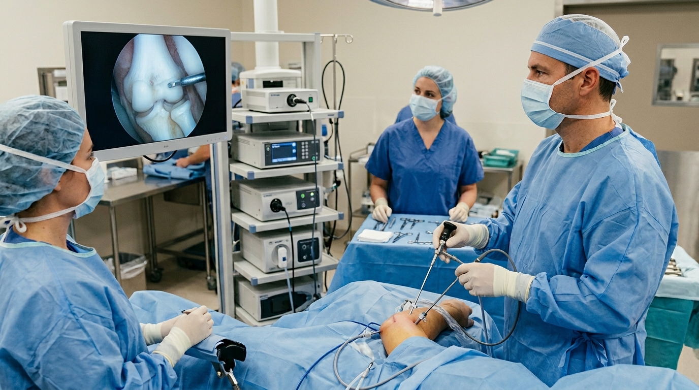
Imaging is where elbow dysplasia is really pinned down. X-rays show arthritis and some of the structural problems, but they frequently miss the detail, particularly a fragmented coronoid process, which often isn't visible on a standard X-ray. CT is much better at identifying the subtle lesions and is commonly used for accurate diagnosis and surgical planning. Arthroscopy, keyhole inspection of the joint, is both the most definitive way to see exactly what's going on inside and, very often, the means of treating it at the same time.
The treatment options
Because elbow dysplasia is several conditions rather than one, the treatment depends heavily on which form your dog has, how severe it is, and the dog's age. I'll describe the broad approaches plainly, but which applies to your dog is firmly a decision for you and a surgeon who has seen the imaging.
Conservative (medical) management
For mild or early cases, and as an entirely reasonable choice for many dogs, conservative management can give good relief. It's the familiar combination: weight control, modified and controlled exercise, pain relief, joint-supporting measures such as omega-3s, and physiotherapy and hydrotherapy.
It's worth knowing that the evidence comparing conservative and surgical management for medial coronoid disease is genuinely mixed, and some studies show comparable outcomes between the two for certain dogs. So conservative management isn't simply the "do less" option; for some dogs it's a legitimate primary choice, and the decision deserves a proper discussion rather than an assumption that surgery is always better.
Surgical management
When surgery is indicated, it's most often done arthroscopically, through small incisions, which allows precise treatment with quicker recovery and less pain than open surgery. What's actually done depends on the specific problem:
- For a fragmented coronoid process or medial coronoid disease, the surgeon typically removes the loose fragment and any damaged cartilage, and cleans up the joint.
- For an ununited anconeal process, options include removing the fragment or, in some young dogs, attempting to reattach it, sometimes combined with a procedure on the ulna to improve the fit.
- For OCD, the loose cartilage flap is usually removed.
- For significant incongruity, procedures that alter the bones to improve the fit may be considered.
Outcomes vary by the specific problem and its severity, and realistic expectations matter here. For medial coronoid disease treated arthroscopically, many dogs improve substantially, though the figures reported across studies vary and some dogs continue to need ongoing management; one body of work suggests a meaningful proportion improve to the point of not needing long-term anti-inflammatories, while severe medial compartment disease carries a more guarded outlook. Ununited anconeal process generally has a less predictable outcome than coronoid disease. The plain summary is that surgery often helps, particularly in removing a clear mechanical problem like a loose fragment, but elbow dysplasia is rarely "cured," and many dogs need continued arthritis management for life regardless of surgery.
Advanced options for severe disease
For severe medial compartment disease where the cartilage is largely worn away, more advanced and specialised procedures exist, including techniques to shift load away from the damaged compartment, and these are very much specialist territory with their own trade-offs. If your dog reaches this point, a specialist will talk you through whether any of these suit your individual case.
What recovery actually involves

Arthroscopic surgery generally involves a shorter, gentler recovery than open or bone-cutting procedures, typically some weeks of restricted, controlled activity with a gradual return to exercise, often supported by physiotherapy and hydrotherapy. More involved procedures, such as those on the ulna or the advanced salvage techniques, carry longer and more demanding recoveries.
Whatever the procedure, the principles from our surgery-decision article apply: controlled rest, a gradual rehabilitation plan, and the discipline to hold your dog back while they heal. And because elbow arthritis usually continues to need management after surgery, recovery here isn't a finish line so much as a step in a longer course of care. The lifelong weight control, appropriate exercise, and the rest of the conservative measures remain central.
Questions worth asking your surgeon
Given the complexity, these questions are particularly useful for elbow dysplasia:
Exactly which form of elbow dysplasia does my dog have, and how severe is it? What does the CT or arthroscopy show? Given that, what are the realistic options, and which do you recommend and why? How much improvement can I realistically expect, and how likely is my dog to need ongoing arthritis management afterwards regardless? Are both elbows affected? What does recovery involve for the procedure you're suggesting? And, because outcomes here are more variable than for some conditions, what does "success" look like in my dog's specific case?
That last question matters especially for the elbow, where "better but still managed" is a common and reasonable outcome, and setting expectations clearly at the outset prevents disappointment later.
The bottom line
Elbow dysplasia is more complicated than most orthopaedic conditions because it's really several conditions under one name, because the imaging and the clinical picture often don't match, and because outcomes are more variable than for, say, a straightforward cruciate repair. None of that means it can't be helped. Removing a loose fragment can make a real difference, conservative management suits many dogs well, and a great deal can be done to keep an affected dog comfortable.
What it does mean is that accurate diagnosis, usually with CT or arthroscopy, and a candid, individualised conversation with a specialist matter even more here than elsewhere. Go in understanding that elbow dysplasia is an umbrella term, that your dog has a specific version of it, that surgery often helps but rarely cures, and that lifelong arthritis management is usually part of the picture. With realistic expectations and good care, most affected dogs can live comfortable, active lives.
Read this alongside the surgery-decision article for the framework on deciding and planning recovery, and lean on the weight, exercise, and therapy articles, because for the elbow, the ongoing conservative management is a central and lifelong part of keeping your dog comfortable.
References
Sister tool · Sightline
Track whether treatment is working
Sightline, a separate ConciergeVet tool, runs a short weekly check-in built on validated pain and mobility instruments, turns it into a single Sightline Score you can watch trend over weeks rather than judge on one bad day, and produces a report you can bring to your vet.
A written log, or our printable trackers, does much the same job.
See how Sightline tracks treatmentSee where your pet stands today
Tracking is the other half of managing arthritis. Take the 2-minute Mobility Check to see your pet's stage, then watch it shift as treatment takes effect.
Take the Mobility CheckYou're not doing this alone
Compare treatment journeys and talk to owners managing arthritis. Free to join.
Join PetsLikeMine