Mast Cell Tumours: The Lump That's "the Great Pretender"
Dr. Alastair Greenway
MRCVS

If your dog's lump has turned out to be, or your vet suspects, a mast cell tumour, you've probably had the same bewildered reaction most owners do. It looked like nothing. A small soft bump, or a little raised patch you assumed was an insect bite or a wart, maybe one that got bigger then smaller and lulled you into leaving it. Hearing that this innocuous thing is cancer feels like a trick.
In a way, it is. Mast cell tumours have a nickname in the profession, "the great pretenders", because they can mimic almost any harmless lump and even change size from week to week (VCA Animal Hospitals). That's the bad news. The good news, and there's real good news here, is that for many dogs a mast cell tumour caught early and removed properly is a cured problem, not a death sentence. Which it turns out to be comes down almost entirely to one word: grade.
What a mast cell tumour actually is
Mast cells are normal cells. They live in your dog's skin and other tissues as part of the immune system, and they're packed with little granules of powerful chemicals, chiefly histamine, the same substance behind an allergic flare-up. Their job is to release those chemicals when needed, to fight parasites and drive inflammation.
A mast cell tumour is what you get when these cells start multiplying out of control, usually forming a lump in or just under the skin. They are the most common malignant skin tumour in dogs, making up somewhere between roughly 7 and 21 percent of all canine skin tumours (VCA Animal Hospitals). Some breeds are more prone than others, with Boxers, Labradors, French Bulldogs, Golden Retrievers and Shar Peis among those most often affected (Śmiech et al., 2019). If your dog is one of these, it's worth being a little more vigilant about new lumps, though it isn't anything you did wrong.
Why they're "the great pretender"
Here's the part that catches everyone out. A mast cell tumour has no single look. It can be a raised pink lump, a soft fatty-feeling bump, a small ulcerated sore, or a patch that resembles an insect bite, a wart or an allergic reaction (VCA Animal Hospitals). And crucially, many of them wax and wane, swelling up one day and settling the next.
That changeability isn't random, and it's the same thing that makes these tumours faintly dangerous to poke. When a mast cell tumour is disturbed, whether by your dog scratching it or by being squeezed, it can "degranulate", dumping its store of histamine into the surrounding tissue. That causes the lump and the skin around it to redden and swell, a reaction vets call Darier's sign (de Nardi et al., 2022). So the lump shrinks back down, you feel reassured, and the cancer carries on. Owners very reasonably read the shrinking as a good sign. It isn't.
Which brings us to the one practical rule worth taking away today: don't keep prodding or squeezing it. Beyond making the local swelling worse, in a minority of dogs a tumour releasing a lot of histamine at once can cause more systemic upset, and ongoing histamine release is also why some dogs with mast cell tumours develop stomach ulcers, with signs like vomiting or a poor appetite (Blackwood et al., 2012). Note where the lump is, photograph it, and leave it alone.
Why grade is the whole story
With most cancers, owners want a single answer to "how bad is it?" With mast cell tumours, that answer almost always waits on the grade, which is the single most important thing your vet will tell you.
Grade describes how aggressive the tumour cells look down the microscope after a lump is removed and sent to a pathologist. There are two grading systems in use. The older Patnaik system sorts tumours into grades 1, 2 and 3, where grade 1 behaves well and grade 3 behaves badly (VCA Animal Hospitals). The newer two-tier Kiupel system, now widely used, simplifies this into just low grade or high grade, partly because the middle Patnaik grade 2 was frustratingly unpredictable (Kiupel et al., 2011).
The difference this makes is enormous, and it's why a generic "mast cell tumour prognosis" is meaningless. Reported average survival is more than two years for low-grade tumours and less than four months for high-grade ones (VCA Animal Hospitals). One study that re-examined those awkward grade 2 tumours under the two-tier system found that the ones reclassified as low grade had a one-year survival of around 94 percent, while those reclassified as high grade sat at about 46 percent (Sabattini et al., 2015). Same word, "cancer". Two completely different roads.
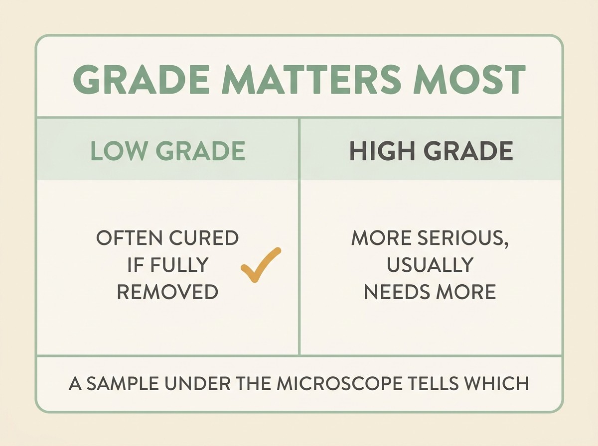
The headline is the reassuring one. The majority of canine mast cell tumours are low grade (Śmiech et al., 2019), and a low-grade tumour that's completely removed is very often a cured dog.
Why "getting it all" matters so much
Surgery is the mainstay of treatment, and with mast cell tumours how the surgery is done genuinely changes the outcome. These tumours send microscopic fingers of cells out beyond what you can feel, so simply shelling out the visible lump tends to leave some behind. That's why surgeons aim for "wide margins", taking a cuff of normal-looking tissue all around and a layer of tissue beneath.
For low-grade tumours under about 4cm, removing the lump with around 2cm of margin to the sides and one tissue layer deep gives a very high chance of clearing it, with reported recurrence rates as low as 0 to 4 percent when the margins come back clean (de Nardi et al., 2022). That is the difference between an operation that fixes the problem and one that leaves it to grow back. You can read more about what "clean margins" means in our guide to cancer surgery.
When a high-grade tumour is found, or the margins come back "dirty", or there are signs it has begun to spread, more is usually needed, and your vet will often recommend a referral to an oncology specialist.
Staging, and treatment beyond the knife
For a small, low-grade tumour that's been cleanly removed, often no further treatment is needed at all, just a check that the local lymph node is clear and an eye kept out for new lumps.
For higher-grade tumours, or ones in awkward spots, the picture gets more involved. Staging means checking whether the cancer has spread, usually by sampling the nearby lymph node and sometimes scanning the tummy (Blackwood et al., 2012). From there the options widen:
- Steroids (prednisolone) can shrink mast cell tumours and calm the histamine effects, and are sometimes used to make a difficult lump smaller before surgery, or as a gentler, comfort-focused option in their own right (Olsen et al., 2018).
- Chemotherapy, often the drug vinblastine combined with prednisolone, is used for high-grade or spreading tumours after surgery. As with all veterinary chemo, the aim is good-quality extra time, and most dogs tolerate it well, which our piece on why pet chemo isn't human chemo explains properly.
- Radiation can mop up tumour left behind when complete surgery isn't possible.
- Toceranib (Palladia) is a tablet, one of a newer class of targeted drugs that block a faulty signal (the KIT protein) driving some of these tumours. In its licensing trial it shrank or stabilised recurrent mast cell tumours in around 37 percent of treated dogs, against about 8 percent on placebo, and dogs whose tumours carry the relevant KIT mutation respond best of all (London et al., 2009).
What to do now, and the one extra check
The most useful things you can do are simple. Don't squeeze or fiddle with the lump. If your dog has the surgery, the result that matters is the grade and whether the margins were clean, so ask for both in plain terms, because they tell you almost everything about what comes next.
And one extra point worth knowing. Most dogs that get a mast cell tumour only ever get the one, but a meaningful minority develop more than one over their life, so finding one is a good reason to keep checking your dog over for new lumps (VCA Animal Hospitals). Our free Lump & Bump Tracker is built for exactly that, letting you log and photograph any new bump so you and your vet can act early, which with this particular cancer is where the good outcomes live.
So hold both truths at once. A mast cell tumour is a real cancer that earned its sneaky reputation. It's also, for the many dogs whose tumour is low grade and fully removed, one of the most genuinely beatable cancers we see. The grade tells you which dog you've got, and that's the number to ask for.
If you want to understand the wider picture, our guide to what grade and stage really mean puts this in context, and if you've found another lump, start with our piece on the lump you've found.
References
- VCA Animal Hospitals. Mast Cell Tumors in Dogs. mast cell tumours as "the great pretenders" mimicking insect bites/warts/allergic reactions and fluctuating in size; "the most common skin tumor in dogs (7%-21%)"; Patnaik grade I-III with grade I much less aggressive than III; "average survival time with high-grade tumors is less than four months, and with low-grade tumors it is more than two years"; advice to avoid palpating or manipulating the tumour because degranulation is easily triggered by pressure; toceranib phosphate (Palladia) as a targeted therapy; "Most dogs with MCT (approximately 85%) only develop one tumor."
- Śmiech A, Łopuszyński W, Ślaska B, Bulak K, Jasik A. Occurrence and Distribution of Canine Cutaneous Mast Cell Tumour Characteristics Among Predisposed Breeds. Journal of Veterinary Research, 2019;63(1):141-148. predisposed breeds (Boxer, Labrador Retriever, French Bulldog, Golden Retriever and American Staffordshire Terrier mainly affected by low-grade MCTs; Shar-Pei the notable exception, at higher risk of high-grade tumours).
- de Nardi AB, et al. Diagnosis, Prognosis and Treatment of Canine Cutaneous and Subcutaneous Mast Cell Tumors. Cells / PMC, 2022. MCTs as "the most common malignant skin tumor in dogs, corresponding to 11% of skin cancer cases"; Darier's sign from degranulation; recommended 2cm lateral margin and one fascial plane deep for grade 1-2 MCTs under 4cm, with a recurrence rate of 0-4%.
- Kiupel M, et al. Proposal of a 2-Tier Histologic Grading System for Canine Cutaneous Mast Cell Tumors to More Accurately Predict Biological Behavior. Veterinary Pathology, 2011. the two-tier (low grade / high grade) grading system proposed to reduce the prognostic uncertainty of the Patnaik grade 2 category.
- Sabattini S, et al. Histologic Grading of Canine Mast Cell Tumor: Is 2 Better Than 3? Veterinary Pathology, 2015. of Patnaik grade II MCTs, those classed low grade vs high grade on the Kiupel system had approximately 94% vs 46% one-year survival probability.
- London CA, et al. Multi-center, Placebo-controlled, Double-blind, Randomized Study of Oral Toceranib Phosphate (SU11654)... for the Treatment of Dogs with Recurrent... Mast Cell Tumor Following Surgical Excision. Clinical Cancer Research, 2009;15(11):3856-3865. objective response rate 37.2% in toceranib-treated dogs versus 7.9% on placebo; better response in dogs with KIT mutations.
- Olsen JA, et al. Combination vinblastine, prednisolone and toceranib phosphate for treatment of grade II and III mast cell tumours in dogs. Veterinary Medicine and Science, 2018;4(3):237-251. prednisolone's effect on mast cell tumours and its use in combination protocols for higher-grade tumours.
- Blackwood L, Murphy S, Buracco P, et al. European consensus document on mast cell tumours in dogs and cats. Veterinary and Comparative Oncology, 2012;10(3):e1-e29. systemic effects of histamine release including gastrointestinal ulceration, and staging guidance including regional lymph node sampling for higher-risk MCTs.
Sister tool · Sightline
Track quality of life over time
Sightline, a separate ConciergeVet tool, runs a short adaptive weekly assessment with a quality-of-life focus mode built around exactly these frameworks, tracks a single composite score over time so you can see the trend rather than judge a single bad day, and produces a Sightline Report PDF you can bring to your vet.
A written log, or our printable quality-of-life sheet, does much the same job.
See how Sightline tracks quality of lifeFound a lump? Track it, and know when to act
A lump cannot be told apart by look or feel — only your vet sampling it can. The Lump & Bump Tracker records its size and how it changes, flags when it has crossed a line worth a vet visit, and builds a clean history to take in.
Open the Lump & Bump TrackerYou're not doing this alone
Compare treatment journeys and talk to owners managing cancer. Free to join.
Join PetsLikeMine