Gallbladder Mucocoele: The Senior-Dog Surprise on the Scan
Alastair Greenway
BVM&S MRCVS
Your older dog went in for an ultrasound, quite possibly for something else entirely, a lump, a routine senior check, a vague off-colour week, and the report has come back with a phrase you have never heard: gallbladder mucocoele. The scan described it as looking like a kiwi fruit, or used the word "stellate", meaning star-shaped. And then somewhere in the conversation the word "rupture" appeared, and now you are anxious, and your dog is standing in front of you looking perfectly fine.
This is one of the more disorienting findings in the whole of liver and gallbladder medicine, precisely because it so often turns up in a dog who seems well. So let us take it calmly and in order: what a mucocoele actually is, why it favours older dogs and what that says about the rest of your dog's health, how to tell the worrying situations from the watchful ones, and what the treatment involves. There is a genuine decision to be made here, and it deserves a clear head.
What a mucocoele actually is
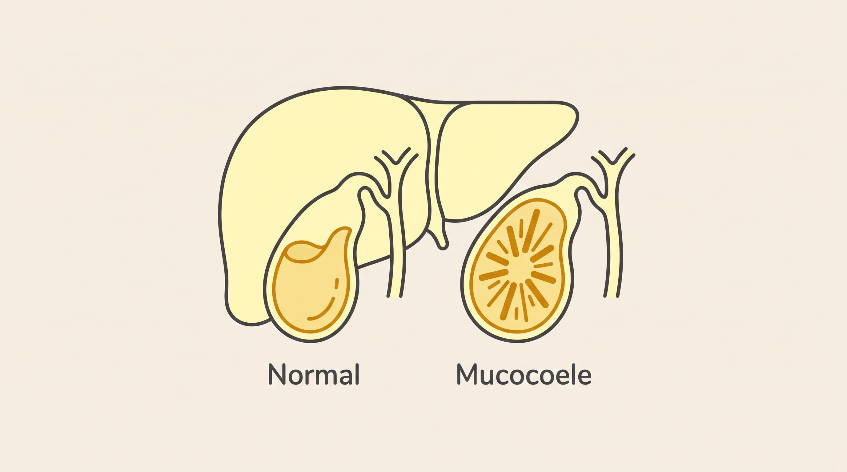
The gallbladder is a small pouch tucked against the liver. Its job is to store bile, the fluid the liver makes to help digest fat, and to squeeze it out into the gut when a meal arrives. Bile is normally a mobile liquid.
In a mucocoele, the gallbladder fills up with thick, gelatinous, immobile bile, more like a firm jelly than a liquid. On ultrasound this sets into a very characteristic pattern, radiating lines from the centre outward, which is why radiologists reach for the "kiwi fruit" or "stellate" description. That distinctive look is often how it gets spotted, sometimes as a complete surprise when the scan was done for another reason.
The reason a mucocoele is not simply a curiosity is twofold. First, that immobile bile can obstruct the normal flow out of the gallbladder. Second, and this is the word that worried you, the distended gallbladder can rupture, spilling bile into the abdomen, which is a surgical emergency and a life-threatening event. Holding both of those facts calmly at once is the task: a mucocoele is a real problem that can become dangerous, and yet many are found in well dogs and handled in a considered way rather than in a panic.
It helps to understand, in plain terms, how bile turns from a free-flowing liquid into a firm jelly. The gallbladder wall normally makes a small amount of mucus, but in a mucocoele the balance tips: the gland produces too much thick mucus, the bile stops moving as it should, and the whole thing sets into that immobile, gelatinous mass. Why that happens is not fully understood in every dog, but abnormal handling of fats and sluggish gallbladder emptying seem central, which is exactly why the hormonal conditions covered below are so tightly linked to it.
Why older dogs, and the endocrine connection
Mucocoeles are overwhelmingly a middle-aged-to-senior dog problem, which is part of why they surface on senior scans. But age is not the whole story, and this is the part that most owners find genuinely useful, because it can uncover a treatable underlying condition.
Mucocoeles are strongly linked to a few hormonal and metabolic conditions. The standout is Cushing's disease (hyperadrenocorticism), where the body produces too much cortisol: dogs with Cushing's have been reported at around 29 times the risk of developing a mucocoele. There are also associations with hypothyroidism (an underactive thyroid) and with high blood fats (hyperlipidaemia). And there is a genetic thread: the ABCB4 gene in Shetland Sheepdogs is a recognised predisposition, which is why Shelties appear disproportionately in the mucocoele literature.
The practical upshot is important. Finding a mucocoele is a good reason to look for one of these underlying conditions, because treating the underlying problem is part of managing the whole dog, and because it may change how your vet thinks about the mucocoele itself. There is a companion piece on the Cushing's, thyroid and gallbladder link that goes into this properly, and it is well worth reading if your dog has a mucocoele or one of these hormonal conditions.
This also reframes the finding in a slightly more hopeful way. A mucocoele can feel like bad luck arriving out of nowhere, but for many dogs it is a clue, a visible sign that something treatable, a hormone imbalance or high blood fats, has been quietly at work. Following that clue can turn a worrying scan into the moment your dog's underlying condition finally gets recognised and managed, which is good for far more than just the gallbladder.
Symptomatic or incidental? This is the fork
Everything about what happens next hinges on one question: is your dog actually unwell, or was this found by chance in a dog who seems fine?
Many mucocoeles are incidental, found in a well dog during a scan for another reason. Others are symptomatic, and the signs to know are vomiting, going off food, lethargy, abdominal pain (a dog who is hunched, tucked-up, or reluctant to be touched around the belly), and jaundice, a yellow tinge to the gums, the whites of the eyes, or the skin. A dog showing those signs is in a different, more urgent situation than a well dog with a chance finding.
That fork, incidental-in-a-well-dog versus symptomatic, is what drives the whole decision, and it is worth stating plainly because it is where owners get frightened unnecessarily or, conversely, too complacent. A ruptured or clearly symptomatic mucocoele is an emergency that usually needs surgery, often urgently. An incidental one in a well dog is a genuine operate-now-versus-monitor conversation, and that decision, which is a real weighing of risks and not a simple yes or no, has its own dedicated piece: mucocoele, operate or watch.
One point worth knowing is that the blood tests can be reassuringly normal, or only mildly abnormal, in a dog with a mucocoele, especially an incidental one. A raised ALP or raised bilirubin can appear as things progress, but you should not take a "normal" liver panel as proof that the gallbladder is fine, because the ultrasound is what actually shows the mucocoele. This is one of the situations where the picture on the scan matters more than the numbers on the printout, and it is why the finding often surprises owners whose dog's bloods looked acceptable.
What surgery involves, and why timing matters
The definitive treatment for a mucocoele is surgical removal of the gallbladder, an operation called a cholecystectomy. Dogs manage perfectly well without a gallbladder afterwards, because the liver still makes bile; it simply flows more continuously into the gut rather than being stored and released in a squeeze.
The crucial point about timing is the gap between planned and emergency surgery. When a cholecystectomy is done electively, on a stable dog before any rupture, outcomes are generally good. When it is done as an emergency after the gallbladder has already ruptured, the dog is far sicker, there is bile contaminating the abdomen, and the risk is considerably higher. This difference is the core of the argument some surgeons make for not waiting once a mucocoele reaches a certain point: the operation you choose calmly is safer than the one forced on you in a crisis. The current thinking on mucocoele management is set out in an ACVS owner overview and a 2025 management review in JAVMA, and your vet or a referral surgeon can place your individual dog against that picture.
None of that means every incidental mucocoele must be whipped out immediately. It means the operate-versus-monitor decision genuinely matters and should be made deliberately, with the rupture risk clearly on the table, which is exactly why it has its own piece.
A word on what to expect after a cholecystectomy, since it is often the owner's next worry. Dogs live perfectly normal lives without a gallbladder; the liver keeps making bile, which simply drips more continuously into the gut rather than being stored and released in a squeeze. Most dogs who come through an elective operation and recover well go on to do very well, which is the whole point of catching a mucocoele before it becomes a crisis. Recovery involves the usual post-abdominal-surgery care, restricted activity and wound checks, and your vet will guide the specifics.
Where this leaves you and your dog
If you have just had this finding land on you, here is a sensible next-steps list. First, if your dog is currently unwell, vomiting, off food, painful, or looking yellow, this is not a "book something for next week" situation; contact your vet promptly, because a symptomatic mucocoele can be the prelude to a rupture. Second, if your dog is well and this was a chance finding, do not panic-book anything at 11pm, but do treat it seriously: ask your vet about screening for the underlying endocrine conditions, Cushing's above all, and about the monitoring plan if you and your vet decide to watch it for now.
And carry this one safety line clearly. A dog with a known mucocoele who becomes suddenly unwell, vomiting, collapsing, in obvious abdominal pain, or jaundiced, may have ruptured and needs an emergency vet immediately. Watching and waiting is a legitimate, monitored choice, but it is never a risk-free one, and knowing the emergency signs is the price of choosing it.
The two pieces to read next are the deeper dive on the operate-or-watch decision, which lays out how the risks are actually weighed, and the piece on the Cushing's, thyroid and gallbladder link, which explains why your dog developed this in the first place and what treating the underlying cause can do. If you decide to monitor, the plan will involve repeat ultrasound scans over time, and keeping to that recheck cadence is how you make watch-and-wait a safe choice rather than a hopeful one.
References
- Webster CRL, et al. ACVIM consensus statement on the diagnosis and treatment of chronic hepatitis in dogs. *J Vet Intern Med* 2019;33(3):1173–1200.
Keep track of how your pet is doing
The owners who cope best are the ones who notice changes early. A simple health log shows you what is working, and what is not, before the next vet visit.
Start tracking, freeYou're not doing this alone
Compare treatment journeys and talk to owners managing liver & gallbladder. Free to join.
Join PetsLikeMine