Cancer Surgery: What Removing It Involves, and "Getting Clean Margins"
Claire Greenway
BVM&S MRCVS
For a great many solid tumours, surgery is the main event. A lump that can be removed completely, with a clean edge around it, is often the closest thing to a cure that pet cancer offers. That's genuinely good news, and it's worth holding on to when the word "operation" lands on top of the word "cancer".
But "we'll remove it" hides a lot of detail, and the detail is what decides whether the surgery works. Two things matter more than almost anything else: getting a margin of healthy tissue around the tumour, and getting it right the first time. This piece covers what your pet's surgery actually involves, what "clean margins" means and why your vet keeps mentioning it, what recovery looks like at home, and what happens if the news isn't quite as clean as everyone hoped.
Why surgery is the mainstay for many cancers
A tumour you can see and feel is a physical lump of abnormal cells. If all of those cells can be cut out, the cancer in that spot is gone. That simple logic is why surgery sits at the heart of treating many solid cancers in dogs and cats, from skin masses to splenic tumours. It's the one treatment that can remove the disease in a single step rather than holding it back over time.
The catch is that not every cancer behaves like a tidy ball. Many send out microscopic roots into the tissue around them, fingers of cells you can't see, can't feel, and can't tell apart from normal flesh by eye. So a surgeon can't just scoop out the lump. They take a deliberate cuff of normal-looking tissue all the way around and underneath it, on the assumption that some cancer cells are hiding in there. That cuff is what a surgical margin is.
What "margins" really means
When a surgeon plans a cancer operation, they decide in advance how much normal tissue to take around the tumour. The standard veterinary teaching is to "excise a normal tissue margin en bloc with the gross tumour", in one piece, so the lump is never cut into (Orencole & Butler, 2013). How wide that margin needs to be depends on the type of tumour. A benign lump might need only a millimetre or two. A soft tissue sarcoma, which is invasive, is often taken with a margin of around 2 to 3cm of normal tissue laterally and at least one layer of tissue (a fascial plane) beneath (Orencole & Butler, 2013). For a feline injection-site sarcoma, which is notoriously invasive, the recommended margins are wider still, in the region of 3 to 5cm laterally and two fascial planes deep (Phelps et al., 2011).
Surgeons describe the "dose" of surgery in levels, and the words crop up in conversations and reports, so they're worth knowing:
- Intralesional means cutting into the tumour itself and leaving visible disease behind. It's rarely the aim for cancer (Orencole & Butler, 2013).
- Marginal means shelling the lump out just outside its capsule. This is fine for many benign lumps, but for a malignant tumour it tends to leave those microscopic roots behind (Orencole & Butler, 2013).
- Wide means taking the tumour with a planned cuff of normal tissue around and under it, aiming to remove both the visible lump and the invisible satellite cells. This is the usual goal of curative-intent cancer surgery (Orencole & Butler, 2013).
- Radical means removing a whole anatomical compartment, such as amputating a leg for bone cancer or removing the spleen.
The single commonest surgical mistake in cancer is using a "dose" that's too low, taking too little tissue (Orencole & Butler, 2013). It's an understandable instinct to be conservative near a beloved pet, but with cancer, being too cautious is its own risk.
Reading the margin result: "clean" versus "dirty"
After surgery, the lump and its surrounding tissue go to a pathology lab. The pathologist inks the outer surface, slices through it, and checks under the microscope whether any cancer cells reach that inked edge. That's where margins become a result rather than a plan.
A complete (or "clean", or "R0") margin means no tumour cells are sitting at the inked edge, so the cancer was, as far as can be told, fully contained in what was removed. An incomplete (or "dirty", or "R1") margin means tumour cells were found right at the edge, so some may have been left behind (Milovancev & Russell, 2017). Words like "clean", "dirty", "close" and "narrow" aren't perfectly standardised between labs and can mean slightly different things, which is one reason vets are increasingly using the clear-cut R0/R1 system borrowed from human medicine (Jennings, Royal Canin Academy).
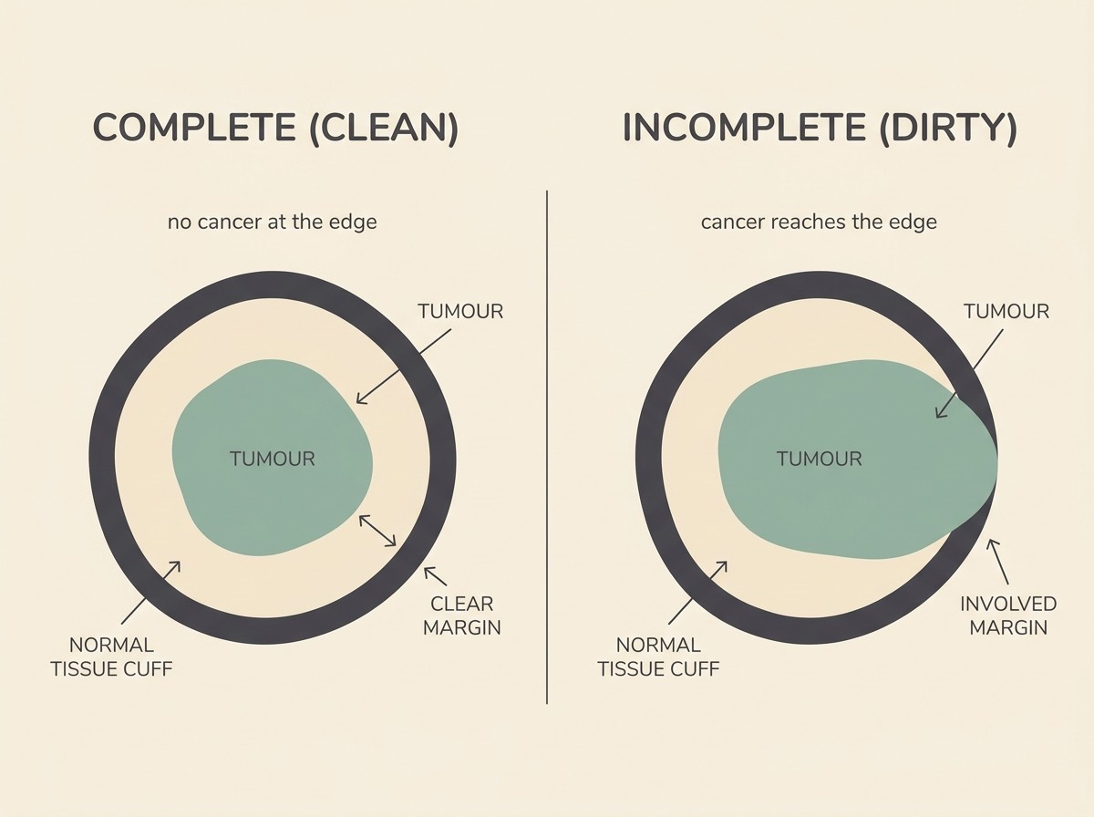
Here's the reassuring part. For many common cancers, clean margins really do mean a very good outcome. With a low-grade mast cell tumour removed with proper margins, recurrence at the site is uncommon, with recent reviews reporting it in only around 0 to 10% of dogs after complete excision (de Nardi et al., 2022). For low-grade tumours like these, the long-term outlook after a clean excision is excellent.
And here's the less-tidy part. An incomplete margin does not mean the cancer will definitely come back. It raises the risk, but plenty of pets with a dirty margin never see a recurrence. Some studies of incompletely excised soft tissue sarcomas, for example, show recurrence in only around a third of cases, and for that tumour the grade matters more than the margin in predicting whether it returns (McSporran, 2009; Jennings, Royal Canin Academy). So a dirty margin is a reason to make a plan, not a reason to despair.
Why the first surgery is the best chance
If there's one idea to take from this whole page, it's this: the first operation is usually the best shot at getting it all. The standard surgical teaching puts it plainly, "the first surgery is often the surgeon's best chance for achieving a positive outcome" (Orencole & Butler, 2013).
The reason is partly mechanical. Once a lump has been operated on, the tissue planes are disturbed, scar tissue forms, and any cancer cells left behind get spread through a wider area. A second operation then has to chase a bigger, blurrier target, and often has to be more aggressive than the first one would have needed to be (Orencole & Butler, 2013). This is why a casual "just whip it off" approach, taking a lump out before anyone knows what it is, can backfire. An unplanned removal of an unknown lump is, in effect, an unplanned marginal excision, and it can make a curable cancer harder to cure (Orencole & Butler, 2013).
It's also the link back to the very start of the journey. The reason we push so hard for a sample before surgery, the aspirate or biopsy in the early stages, is so the surgeon knows what they're dealing with and can plan the right margin the first time rather than discovering the problem after the fact.
Planning the operation: tests first, then surgery
Good cancer surgery rarely starts with a scalpel. It usually starts with information. Knowing the tumour type, and sometimes its grade, tells the surgeon how wide to go (Orencole & Butler, 2013). Staging tests, such as blood work, imaging of the chest or abdomen, and sometimes sampling a nearby lymph node, check whether the cancer has spread, because there's little sense in a big operation on the lump if the disease is already widespread. Sometimes the plan is to biopsy first, get the answer, then operate with the right margins; sometimes, for a small accessible lump where the type is fairly clear, your vet goes straight to a planned wide removal. Either way, the surgery is designed around what the tumour is, not improvised on the day.
What recovery is like at home
For most pets, recovery is more straightforward than owners fear. Your pet usually comes home the same day or the next, groggy from the anaesthetic, and that wears off over about 24 to 48 hours (VCA Animal Hospitals). Any skin stitches are usually removed about 7 to 14 days after the operation, by which point the incision has typically healed over, though deeper or larger wounds take longer (VCA Animal Hospitals).
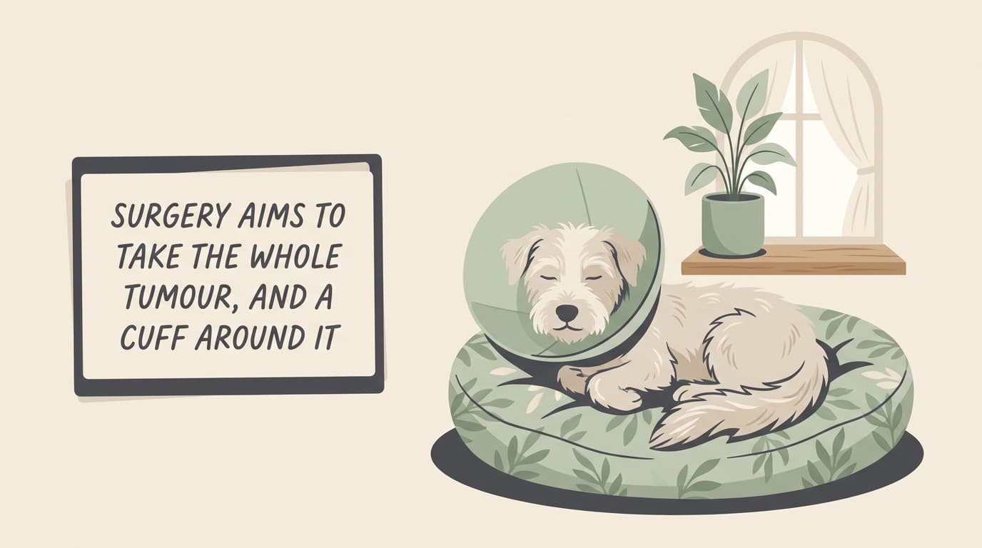
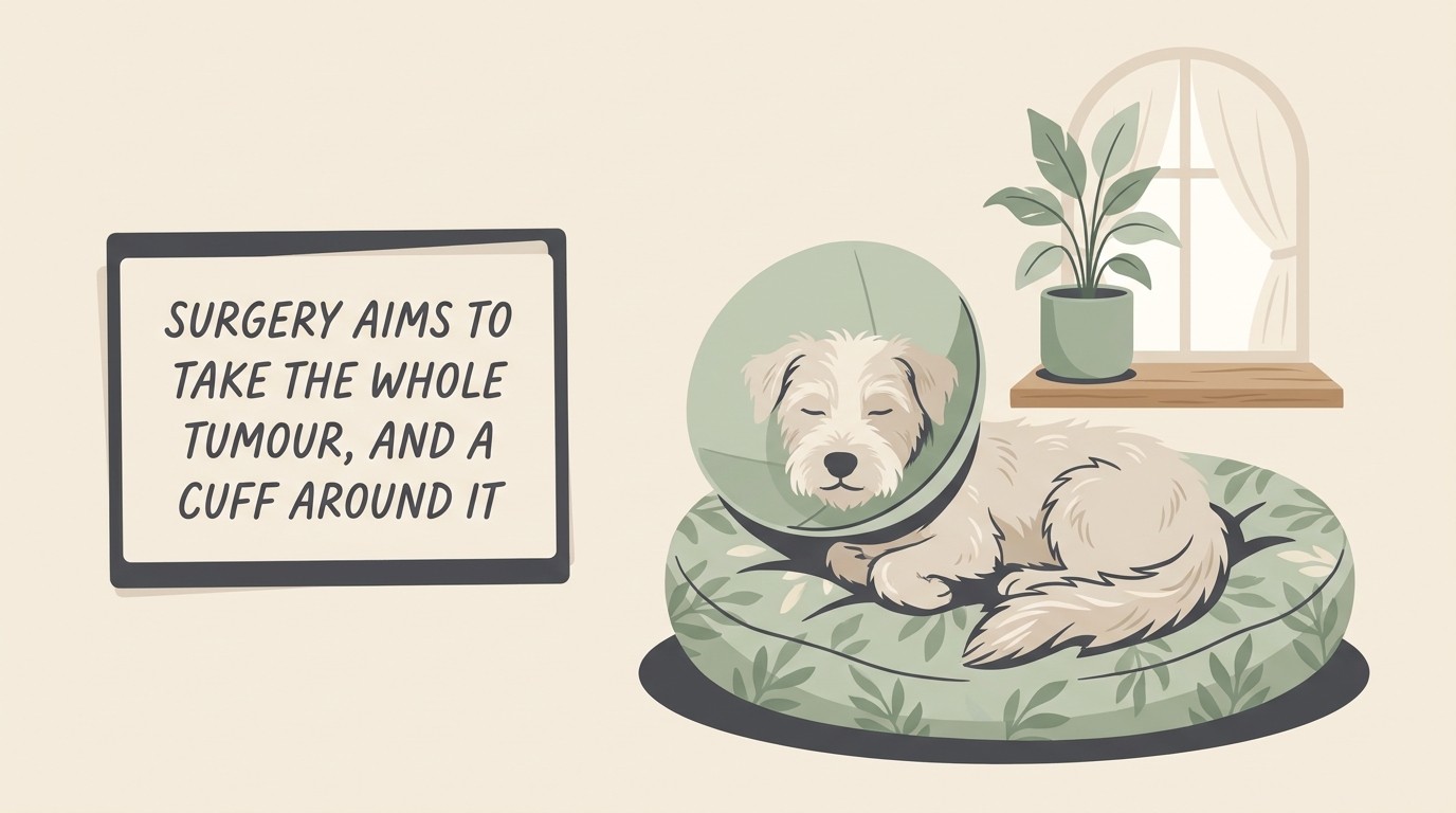
The main jobs at home are calm and protection. Your pet needs genuine rest, usually for around two weeks, with lead walks only, no running, jumping or stairs, and a quiet space to settle. The wound needs to stay clean and dry and to be left alone, which is what the cone (the Elizabethan collar) or a recovery bodysuit is for. Licking is the enemy, because it can open or infect a healing incision, so the cone stays on whenever your pet isn't directly supervised, including overnight, usually for about 10 to 14 days (VCA Animal Hospitals). It looks miserable, but most pets get used to it within a day, and it does its job. You'll be sent home with pain relief, and it's worth checking the wound once or twice a day for the things that mean a phone call: increasing redness, swelling, discharge, a bad smell, or the edges coming apart. A little bruising and a thin line of pinkish skin are normal; an angry, weeping or gaping wound is not.
When surgery isn't the answer
Surgery is powerful, but it isn't always possible or wise. Some tumours sit somewhere that can't be removed without taking out something the body needs, or wrap around vital structures. Some cancers have already spread by the time they're found, so removing the original lump wouldn't change the outcome. And for some pets, the operation or the recovery isn't a fair thing to ask given their age or other health problems.
When that's the case, surgery being off the table doesn't mean help is off the table. If a margin can't be made clean by re-operating, radiation can often mop up the microscopic disease left behind and protect against regrowth. And where surgery isn't right at all, comfort-focused care can keep a pet genuinely well for a long time. None of those is a lesser choice; they're different routes to the same goal of good, comfortable time. There's a money side to all of this too, which is a legitimate part of the decision and not something to feel awkward about. A cancer operation, with the anaesthetic, the pathology and the follow-up, is a real cost, so ask for a written estimate up front, and see our separate piece on what treatment costs and how people manage it.
If your pet is heading for surgery, the most useful thing you can do is ask the two questions that matter most: what type of tumour is this, and what margin are you aiming for. The answers tell you almost everything about what the operation is trying to achieve, and they put you and your vet on the same side of a plan built around your pet.
References
- Orencole, M.J., & Butler, R. (2013). Fundamentals of Surgical Oncology in Small Animals. Today's Veterinary Practice (Nov/Dec 2013).
- Milovancev, M., & Russell, D.S. (2017). Surgical margins in the veterinary cancer patient. Veterinary and Comparative Oncology, 15(4), 1136 to 1157.
- Jennings, R. The Role of Surgical Margins and Histology in Tumor Excision. Royal Canin Academy (veterinary education resource; discusses the R0/R1 classification, the ambiguity of "clean/dirty", and grade versus margin as a predictor in soft tissue sarcoma).
- American Animal Hospital Association (2026). 2026 AAHA Oncology Guidelines for Dogs and Cats (clean surgical margins as the key factor in preventing local recurrence).
- VCA Animal Hospitals. Post-Operative Instructions in Dogs (recovery timeline, incision care, Elizabethan collar use and suture removal).
- Haine, M., et al. (2022). Incomplete histological margins following planned narrow excision of canine appendicular soft tissue sarcomas and mast cell tumors, using the residual tumor classification scheme. Veterinary Surgery, 51(4). (supports the R0/R1 "tumour on ink" definition of incomplete margins).
- Phelps, H.A., Kuntz, C.A., Milner, R.J., et al. (2011). Radical excision with five-centimeter margins for treatment of feline injection-site sarcomas: 91 cases (1998 to 2002). Journal of the American Veterinary Medical Association, 239(1), 97 to 106.
- de Nardi, A.B., et al. (2022). Diagnosis, Prognosis, and Treatment of Canine Cutaneous and Subcutaneous Mast Cell Tumors. Cells, 11(4), 618.
- McSporran, K.D. (2009). Histologic grade predicts recurrence for marginally excised canine subcutaneous soft tissue sarcomas. Veterinary Pathology, 46(5), 928 to 933.
Sister tool · Sightline
Track quality of life over time
Sightline, a separate ConciergeVet tool, runs a short adaptive weekly assessment with a quality-of-life focus mode built around exactly these frameworks, tracks a single composite score over time so you can see the trend rather than judge a single bad day, and produces a Sightline Report PDF you can bring to your vet.
A written log, or our printable quality-of-life sheet, does much the same job.
See how Sightline tracks quality of lifeFound a lump? Track it, and know when to act
A lump cannot be told apart by look or feel — only your vet sampling it can. The Lump & Bump Tracker records its size and how it changes, flags when it has crossed a line worth a vet visit, and builds a clean history to take in.
Open the Lump & Bump TrackerYou're not doing this alone
Compare treatment journeys and talk to owners managing cancer. Free to join.
Join PetsLikeMine