"My Vet Wants to Take a Sample": Aspirates, Biopsies and What the Results Mean
Claire Greenway
BVM&S MRCVS
Your vet has felt the lump, looked thoughtful, and said the thing you didn't quite want to hear: "I'd like to take a sample." It's a small sentence that can set off a big worry. Does wanting a sample mean they think it's cancer? Will it hurt? How long will you have to wait, and what on earth will the result actually say?
So here's the reassuring shape of it before we get into the detail. Taking a sample is the most normal, sensible next step after finding a lump, and it's exactly what we'd want for our own pets. It's how a guess becomes an answer. The commonest test is quick, usually done with your pet wide awake, and most dogs and cats barely notice it. And the result, once it comes, is far less mysterious than it sounds. This page walks you through the two main tests, what each can and can't tell you, the wait, and how to read the report in plain English.
If you've landed here straight from finding a lump, our guide on the lump you've found explains why getting it sampled beats watching and waiting in the first place.
The two tests, and why they're different
There are two main ways to look at what a lump is made of, and they answer slightly different questions.
The first is a fine needle aspirate, usually shortened to FNA. Your vet puts a small needle, often no bigger than a vaccine needle, into the lump and collects a few cells, either by gentle suction or a little "pecking" motion. Those cells are squirted onto a glass slide, spread thin, stained and examined under a microscope. Looking at loose cells like this is called cytology. It's the everyday first-line test for a skin or subcutaneous lump, and it's an excellent screening tool (Today's Veterinary Practice).
The second is a biopsy, where instead of a few cells your vet takes an actual piece of tissue, either a small core or, sometimes, the whole lump. That tissue is processed, sliced wafer-thin and examined intact. Looking at tissue with its structure preserved is called histopathology. Because the architecture is kept, a biopsy can see things cytology can't, which is why it's the more definitive of the two (Today's Veterinary Practice).
The simplest way to hold the difference in your head: an aspirate looks at the cells, a biopsy looks at the tissue. One is a quick peek, the other is the full read.
The needle test, from your pet's point of view
This is the bit owners worry about most, and it's usually the least dramatic part of the whole thing.
A fine needle aspirate is quick, often over in a minute or two, and for an accessible skin lump it almost always needs no sedation at all. Most pets sit for it the way they'd sit for a vaccination, and the discomfort is on a par with exactly that, a quick small scratch (DogCancer, 2023). There's no cut, no stitches and nothing to recover from afterwards. Your dog can have a treat and go home, or your cat can go straight back to pretending the whole visit never happened.
A biopsy is a bigger undertaking, because taking a piece of tissue does need your pet to be still and comfortable. That usually means sedation or a short general anaesthetic, and sometimes a stitch or two afterwards (Today's Veterinary Practice). It's still a routine procedure, but it's a planned one rather than something done casually in the consult room, which is part of why vets often reach for the aspirate first.
What each test can, and can't, tell you
Here's where it helps to be plain about the strengths and the limits, because it explains why your vet might choose one route over the other.
A fine needle aspirate is brilliant at answering the big first question, "what kind of thing is this?". For lumps in and under the skin, cytology agrees with the final tissue diagnosis remarkably often. In one study of cutaneous and subcutaneous masses in dogs and cats, the cytology matched the histopathology in around 91% of cases, and it was very good at picking up cancer when it was there (Ghisleni et al., 2006). For many lumps, that quick needle test gives you a clear, trustworthy answer the same day or within a few days.
But an aspirate has two real limitations, and they're worth knowing so a result doesn't throw you.
The first is that it sometimes comes back non-diagnostic, which simply means not enough usable cells came out to say anything for certain. It is not a hidden bad sign and it is not anyone's fault. Some lumps just don't give up their cells easily. In that same skin-mass study, around one in six samples didn't have enough cells to read (Ghisleni et al., 2006), and for certain sites the rate is higher still, with lymph node aspirates non-diagnostic in roughly a quarter of dogs (27%) and around one in seven cats (14%) in one large study (Amores-Fuster et al., 2015). The fix is straightforward: repeat the aspirate, or move on to a biopsy.
The second limitation is that an aspirate often can't grade a cancer, and for some tumours it can't fully pin down the type. Dense tumours like soft tissue sarcomas are notorious for not shedding cells well, so they frequently need a tissue biopsy to be sure (DogCancer, 2023; Today's Veterinary Practice). Grading, how aggressive a cancer looks down the microscope, almost always needs a proper tissue sample. We explain why grade and stage matter so much in our guide to what those words mean.
That's the whole logic of it. The aspirate is the fast, gentle screen that's right most of the time, and the biopsy is the fuller answer when the screen isn't enough or when your vet needs the grade and the detail to plan properly. Neither is "better"; they do different jobs.
Why your vet might go straight to a biopsy
Sometimes your vet will skip the needle test and recommend a biopsy from the off, and that isn't a sign they're expecting the worst. It usually just means a biopsy is the more sensible first step for that particular lump.
That might be because of where the lump is, or its size, or the type they suspect from experience, or because the plan is to grade it accurately before deciding on surgery. For a lump that's clearly going to be removed whatever the cytology shows, it can make sense to take it out and send the whole thing to the lab in one go. None of this is a verdict. It's your vet matching the test to the job.
The wait, and what's happening to the sample
The wait for results is often the hardest part, so it helps to know what's going on while you're refreshing your phone.
A sample doesn't sit in a drawer. It's sent to a veterinary laboratory where a specialist pathologist processes it, examines it and writes a report. A straightforward skin cytology can sometimes be read quickly, occasionally even in-house the same day. A tissue biopsy takes longer, because the tissue has to be carefully prepared before it can be cut and stained. As a rough guide, one of the large veterinary labs quotes around three to five days for a standard biopsy, with more complex specimens, or those needing extra processing, taking longer, commonly five to seven days and occasionally more (IDEXX Reference Laboratories).
If the wait stretches, it's almost always because the lab is being thorough rather than because something alarming has turned up. A pathologist taking an extra day to run a special stain is doing exactly what you'd want.
Reading the report in plain English
When the report lands, it can look like a wall of unfamiliar words. It's actually answering a short list of practical questions, and once you know what they are it reads far more calmly.
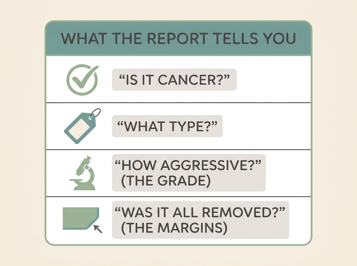
- Is it cancer at all? The first and biggest answer. Plenty of reports come back describing a fatty lump (a lipoma), a cyst or another harmless swelling, and that's the end of the story.
- If so, what type? Cancer is dozens of different diseases, and the name matters far more than the word "cancer" on its own. The type is what tells your vet, and you, what you're actually dealing with.
- How aggressive does it look (the grade)? Usually only on a tissue biopsy. Low grade tends to mean slower and less likely to spread; high grade means the opposite. Grade shapes the whole plan.
- Was it all removed (the margins)? Only relevant when a lump has been taken out. The pathologist checks the edges of what was removed, and the large majority of reports describe this (around 92% in one US review of mast cell tumour reports; Reagan et al., 2018). "Clean" or complete margins mean a clear rim of normal tissue all the way round; "dirty" or incomplete margins suggest some microscopic cancer may have been left, which simply means a decision about whether more is needed. Our guide to cancer surgery and clean margins covers what happens then.
Put those four answers together and you have the foundation of everything that comes next: whether there's anything to treat, and if so, what the realistic options are. That's exactly the picture our guide on what grade, stage and prognosis really mean helps you build.
What "inconclusive" means, and what comes after
If your result is non-diagnostic or inconclusive, try not to read anything into it beyond the plain meaning: the test didn't gather quite enough to be sure. As above, it happens to a fair share of perfectly ordinary lumps, and it's not a clue that something is being hidden from you.
The next step is usually simple. Your vet may repeat the aspirate, often it works the second time, or move up to a biopsy for the fuller tissue answer. Occasionally the most efficient plan is to remove the lump and let the lab examine the whole thing, which gives you the type, the grade and the margins all at once.
A quick word on cost, since it's a fair question to ask out loud. A fine needle aspirate is the more affordable test, which is another reason it's the usual first move; a biopsy with a full histopathology report costs more because there's more involved, from the anaesthetic to the lab work. Prices vary between practices and labs, so it's completely reasonable to ask for an estimate before you go ahead, and your vet would far rather you asked than worried about it silently.
Whatever route your lump takes, the sample is the thing that turns fear of the unknown into a clear picture you can act on. The needle test is usually quick and easily shrugged off, the biopsy is routine, and the report, read through those four questions, is far less frightening than the wait makes it feel. When the result comes back, our guide on what grade, stage and prognosis actually mean is the natural next read.
References
- Ghisleni G, Roccabianca P, Ceruti R, Stefanello D, Bertazzolo W, Bonfanti U, Caniatti M. Correlation between fine-needle aspiration cytology and histopathology in the evaluation of cutaneous and subcutaneous masses from dogs and cats. Veterinary Clinical Pathology. 2006;35(1):24-30. DOI: 10.1111/j.1939-165X.2006.tb00084.x. (Cytology agreed with histopathology in 90.9% of cases; ~16.8% of samples non-diagnostic due to poor cellularity; sensitivity 89.3%, specificity 97.9% for neoplasia.)
- Today's Veterinary Practice. Ultrasound-Guided Fine Needle Aspiration and Core Biopsy. (FNA yields cells for cytology and usually needs little to no sedation; core biopsy yields tissue for histopathology with architectural assessment and needs heavy sedation or anaesthesia; cytology can be non-specific or fail to correlate with histopathology.)
- Lester KM (reviewed by Ubell A). Fine Needle Aspiration for Dogs. DogCancer.com, updated 2023. (FNA usually needs no sedation for skin masses, most dogs barely feel the needle, discomfort comparable to a vaccine; some tumours such as sarcomas do not aspirate well and need a biopsy.)
- Christensen J, Johnson K, Ettinger S, Garrett L, Gordon I, Ireifej S, Love A, Wisecup M. AAHA Oncology Guidelines for Dogs and Cats. Journal of the American Animal Hospital Association. 2026;62(1):1-37. DOI: 10.5326/JAAHA-MS-7549. (Treatment is built on first identifying the tumour's type, grade and stage.)
- Ettinger SN. A Practical Approach to Lumps and Bumps. WSAVA World Congress Proceedings, 2017. (Via Veterinary Information Network.) ("See something, do something"; a mass over ~1cm or present a month should be aspirated or biopsied rather than watched.)
- Amores-Fuster I, Cripps P, Graham P, Marrington AM, Blackwood L. The diagnostic utility of lymph node cytology samples in dogs and cats. Journal of Small Animal Practice. 2015;56(2):125-130. DOI: 10.1111/jsap.12303. (Of 1274 canine lymph node samples, 27.2% were non-diagnostic; of 199 feline samples, 14.1% were non-diagnostic.)
- IDEXX Reference Laboratories. Histopathology Services. (Standard biopsy results in 3-5 days; complex biopsies in 5-14 days, typically final within 7 unless extended processing is required, e.g. for bone.)
- Reagan JK, Selmic LE, Fallon C, Driskell EA, Garrett LD. Evaluation of information presented within mast cell tumour histopathology reports in the United States: 2012-2015. Veterinary Medicine and Science. 2018;4(4):311-321. DOI: 10.1002/vms3.107. (Surgical margins were reported in 92% of histopathology reports.)
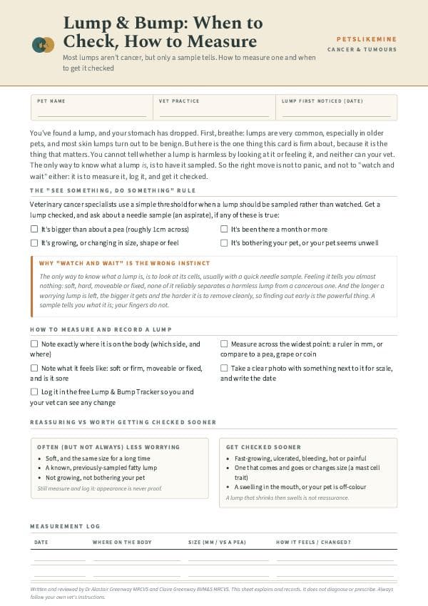
Free downloads
Companion worksheets to put what you've read into practice. Free PDFs, print at home.
Sister tool · Sightline
Track quality of life over time
Sightline, a separate ConciergeVet tool, runs a short adaptive weekly assessment with a quality-of-life focus mode built around exactly these frameworks, tracks a single composite score over time so you can see the trend rather than judge a single bad day, and produces a Sightline Report PDF you can bring to your vet.
A written log, or our printable quality-of-life sheet, does much the same job.
See how Sightline tracks quality of lifeFound a lump? Track it, and know when to act
A lump cannot be told apart by look or feel — only your vet sampling it can. The Lump & Bump Tracker records its size and how it changes, flags when it has crossed a line worth a vet visit, and builds a clean history to take in.
Open the Lump & Bump TrackerYou're not doing this alone
Compare treatment journeys and talk to owners managing cancer. Free to join.
Join PetsLikeMine